Diagnosis of Apoptosis – Lab Techniques and Biomarkers
“What is apoptosis and how is it diagnosed?”
Apoptosis is a form of coordinated and internally programmed cell death.
Apoptosis Morphologic Changes
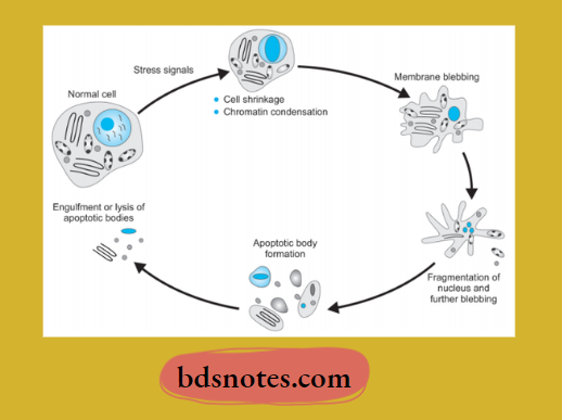
Following are the morphological changes in apoptosis which are seen in an electron microscope:
- Cell shrinkage: The cell is smaller in size; the cytoplasm is dense, and the organelles, although relatively normal, are more tightly packed.
- Chromatin condensation: This is the most characteristic feature of apoptosis. The chromatin aggregates peripherally, under the nuclear membrane, into well-delimited dense masses of various shapes and sizes. The nucleus itself may break up, producing two or more fragments.
- Formation of cytoplasmic blebs and apoptotic bodies: The apoptotic cell first shows extensive surface blebbing, then undergoes fragmentation into several membrane-bound apoptotic bodies composed of cytoplasm and tightly packed organelles, with or without a nuclear fragment.
- Phagocytosis of apoptotic cells or bodies by adjacent healthy cells, either parenchymal cells or macrophages. The apoptotic bodies are rapidly degraded within lysosomes, and the adjacent cells migrate or proliferate to replace the space occupied by the now-deleted apoptotic cell.
Apoptosis diagnosis
“Global prevalence of apoptosis-related diseases”
“Understanding apoptosis: Causes and diagnostic methods”
Molecular Mechanism Of Apoptosis
1. Initiators Of Apoptosis
- Stimulation of signaling programmed cell death acts either at the cell membrane or intracellularly. This includes:
- Absence of stimuli required for normal cell survival.
- Activators of programmed cell death.
- Intracellular stimuli include heat, radiation, hypoxia, etc.
Apoptosis biomarkers
“Importance of diagnosing apoptosis in disease research”
2. Regulators of apoptosis
- As the cell is involved in apoptosis by the above-mentioned signals, the next phase is regulation, in which certain proteins convert death signals to fail programmed cell death and determine the outcome. Regulator proteins are:
- BCL-2: It is a gene that is located in the outer mitochondrial membrane and may regulate the apoptotic process by binding to some other proteins, For Example. As BCL-2 binds to BAX or A, D, it promotes apoptosis, while as it binds to BCL-XL, it inhibits apoptosis.
- Other apoptotic regulator proteins: Besides BCL-2, other regulatory proteins of apoptosis are TP53 protein, BAX, and certain viruses.
“Case studies on outcomes of apoptosis diagnosis”
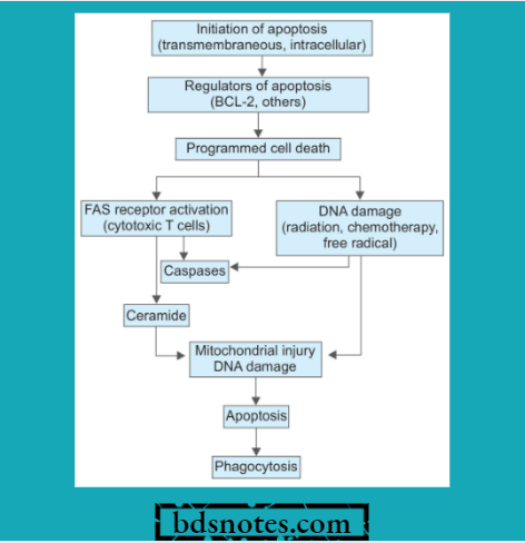
3. Programmed Cell Death
- The outcome of apoptotic regulators in programmed cell death involves the following pathways:
- FAS receptor activation: Cell surface receptor FAS is present in cytotoxic T cells. On coming in contact with target cells, the FAS receptor is activated. This activates caspases and leads to proteolysis and further apoptosis.
- Ceramide generation: Due to the hydrolysis of a phospholipid, i.e., ssphingomyelinasen of the plasma membrane,e ceramide gets generated. Ceramide is implicated in mitochondrial injury and further apoptosis.
- DNA damage: Damage to DNA by ionizing radiation, chemotherapeutic agents, and activation of oxygen species leads to apoptosis. DNA damage affects TP,53, which induces the synthesis of cell death-promoting protein BAX and further apoptosis.
- Phagocytosis: Dead apoptotic cells and their fragments possess cell surface receptors that facilitate their identification by adjacent phagocytes. Phagocytes engulf the apoptotic cells and clear the area.
Assays for apoptosis
“Impact of flow cytometry on apoptosis analysis”
Diagnosis Of Apoptosis
- Agarose gel electrophoresis shows a step ladder pattern
- Terminal deoxynucleotidyl transferase biotin-dUTP nick end labeling (TUNEL) technique for in vivo detection
- H&E stain, Feulgen, and acridine orange staining of apoptotic cells
- Measurement of cytosolic cytochrome C and activated caspase
- Expression of phosphatidyl serine on the outer leaflet of the plasma membrane by apoptotic cells enables their recognition by using the dye annexin V.
“Emerging research on apoptosis detection techniques”
Disorders Associated With Apoptosis
- Disorders associated with decreased apoptosis, i.e., cancer, autoimmunity
- Disorders associated with increased apoptosis:
- Neurodegenerative diseases, i.e., Alzheimer’s Huntington’son, Parkinson’s disease
- Ischemic injury in stroke and myocardial infarction
- Death of virus-infected cells as in AIDS.
Leave a Reply