Development Of Tooth
Give the microscopic structure of Developing tooth Collagen Fibres Golgi apparatus
Answer:
Developing tooth Collagen fibres Golgi apparatus Developing tooth:
- The epithelium in relation to the alveolar process thickens to form dental lamina
- The cells of dental lamina proliferate at various sites to form enamel organ
- This enamel organ grows into underlying mesenchyme & assumes cup shaped appearance
- The mesenchyme is of neural crest origin & is called dental papilla
- The dental papilla alongwith enamel organ is called tooth germ
- This stage is called ‘cap stage’
- The cells of enamel organ adjacent to dental papilla becomes columnar & are known as ameloblasts
“Factors influencing success with tooth studies: Q&A”
- The mesenchymal cells adjacent to ameloblasts differentiate into odontoblasts
- The two cell layers are separated by a basement membrane
- Rest of mesenchymal cells form pulp
- This is called ‘bell stage’
- Ameloblasts lay down enamel & odontoblasts lay down dentin
- The root of the tooth is formed by laying down of layers of dentin & narrowing the pulp space
- The root dentin is covered by mesenchymal cells that differentiate into cementoblasts
- These cementoblasts lay down cementum
“Understanding tooth development through FAQs: Stages, functions, and uses explained”
Developing tooth Collagen fibres Golgi apparatus Collagen fibres:
- Each collagen fibre is made up of fibrils of diameter 20200 nm
- Each fibrils shows cross striations
- Each fibrils contains numerous myofibrils
- Size of each collagen fibre is about 112 micrometer in diameter They are arranged in groups
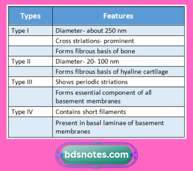
Developing tooth Collagen fibres Golgi apparatus Types:
“Importance of studying tooth development for dental students: Questions explained”
Developing tooth Collagen fibres Golgi apparatus Golgi apparatus:
- It is small, irregular structure
Developing tooth Collagen fibres Golgi apparatus Location:
- Usually near the nucleus
Developing tooth Collagen fibres Golgi apparatus Structure:
- It is made up of membranes
- Membranes form the walls of a number of flattened sacs that are placed over one another
- Towards the margins there are small rounded vesicles
“Common challenges in mastering tooth notes effectively: FAQs provided”
Developing tooth Collagen fibres Golgi apparatus Regions:
- Functionally, Golgi apparatus is divided into 3 regions
1. Cis face/ Cis Golgi
- It is a region near to the nucleus
2. Trans face/ Trans Golgi
- It is a region near cell membrane
3. Intermediate part
- It is present between cis & trans face
Leave a Reply