Dental Implants
Question 1. Write short note on osseointegration.
Or
Describe osseointegration.
Or
Write short answer on osseointegration.
Answer.
Osseointegration Introduction or Osseointegration Definition
Osseointegration Of Dental Implants
- In the 1950s, Per-Ingvar Brånemark, a Swedish professor of anatomy, had a serendipitous finding while studying blood circulation in bone that became a historical breakthrough in medicine. He discovered an intimate bone-to-implant apposition with titanium that offered sufficient strength to cope with load transfer. He called the phenomenon osseointegration and developed an implant system with a specific protocol to predictably achieve it.
- Histologically, osseointegration is defined as the direct structural and functional connection between ordered, living bone and the surface of a load-bearing implant without intervening soft tissues.
- Clinically, osseointegration is the rigid fixation of an alloplastic material (implant) in bone with the ability to withstand occlusal forces.
- Branemark also stated that the implant should not be loaded during healing period for osseous integration to occur.
- Osseointegration is a clinically asymptomatic rigid fixation of the implant within bone, during functional loading.
- There is no connective tissue intervening between bone and implant so the interface is strong one which can withstand the occlusal loads.
- Interface consists of remodeled bony tissue. To develop this strong interface, the implant should not be overloaded during its organization period (Soon after placement of implant).
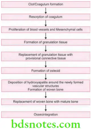
- During this period, the surgical area undergoes a remodeling process just like an extraction site.
- During organization the bone grows into the irregularities of the implant surface
- Hydroxyapatite coated implant get “Biointegrated” with the bone tissue.
Read And Learn More: Periodontics Question And Answers
Basic Principles of Implant Therapy to Achieve Osseointegration
- Implants must be sterile and made of a biocompatible material (e.g., titanium).
- Implant site should be prepared under sterile conditions.
- Implant site should be prepared with an atraumatic surgical technique that avoids overheating of the bone during preparation of the recipient site.
- Implants should be placed with good initial stability.
- Implants should be allowed to heal without loading or micromovement (i.e., undisturbed healing period to allow for osseointegration) for 2 to 4 or 4 to 6 months, depending on the bone density, bone maturation, and implant stability.
Stages of Osseointegration or Osseointegration Process
Factor Affecting Osseointegration
Overload: It causes failure of osseointegration. Premature loading of implant at the time of healing leads to fibrous encapsulation and not osseointegration.
- Biocompatibility of the material.
- Implant design: Most conductive design for osseointegration is cylindrical.
- Implant surface: Implant with mild surface roughness leads to more osseointegration.
- Surgical site: A healthy site is required previously irradiated area is contraindicated.
- Surgical technique: Minimum possible trauma. Surgical drilling should be intermittent, slow rate and is by using sharp instrument.
- Infection control: Infection especially form the periodontium should be avoided.
Question 2. Write short note on dental implant.
Answer. Dental implant is an integral component of oral implant complex which also consists of supportive bone, interposed keratinized and mucosal oral soft tissues and prosthetic suprastructure.
Structures of an Osseointegration Implant System
It consists of two types of structures, i.e.
- Implant
- Endostructure or primary element or implant
- Superstructure
- Mesostructure or secondary element or abutment
- Exostructure or tertiary element or superstructure
Endostructure or Primary Element or Implant Osseointegration
Osseointegration Implant Apex
The implant apex is the lower (apical) part of the implant body, through which the vertical force exerted on the implant is transmitted to the jaw bone. Screw implants transmit the vertical force via the thread into the bone.
Osseointegration Implant Body
The part of a root replacement that is positioned in the bone (intraossal) is known as implant body. The coated, perforated implant body is of two types, i.e. hollow body and solid body.
Osseointegration Implant Neck
The implant neck is located between the implant body in the jaw bone and the implant shoulder. An implant neck with a machine treated surface prevents plaque accumulation. Given the subgingival placement of the implant neck, the mucous membrane may adapt without irritation. Implants inserted into the alveolar ridge do not require a pronounced implant neck.
Osseointegration Implant Shoulder
The implant shoulder is the transition between the implant neck and the implant post. The implant shoulder is narrow with a machine treated surface and may be bevelled to improve the aesthetic appearance.
Osseointegration Implant Head
The implant head is the most coronal part of the implant and it represents the connection to the implant post or directly to the superstructure. There are implant heads with antirotation (for single crowns) and without antirotation lock (for bridges). If an antirotation lock is present, it can be integrated within or outside the implant head.
Implant Osseointegration Superstructure
It consists of every component which is retained by the implant and protrudes in oral cavity. These are secondary elements and tertiary elements:
Osseointegration Dental – Mesostructure or Secondary Element
- Abutment: The abutment is the part of a one or two phase implant system which is connected to the implant or fixed to it. It is the build-up that protrudes into the oral cavity, which is either directly included into the superstructure or which serves as a connection element between the implant and the superstructure.
- Implant abutment screw: Implant screw also called abutment screw, is used for a rigid, mechanically stable screw connection between the implant, abutment, and superstructure.
Osseointegration Dental – Tertiary Element or Exostructure
- Superstructure: The superstructure is the prosthetic restoration that is either directly or, in most cases, indirectly connected with the implant. It may be retained on implants and natural abutment teeth at the same time. Depending on the type of connection, superstructures are classified into fixed, partly removable, and removable superstructures.
- Horizontal transocclusal screw: With this screw, superstructures are screwed down transocclusally or horizontally to form partly removable structures.
Osseointegration Of Dental Implants – Classification of Osseointegration Implants
- Based on shape and position in jaws
- Subperiosteal implant
- Transosteal implant
- Endosseous implant
- According to the body shapes
- Threaded implants
- Threadless/Smooth implants
- According to surface characteristics
- Additive surface treatment
- Titanium plasma spraying
- Hydroxyapatite-coating surface
- Substractive surface treatment
- Blasting with titanium oxide/aluminum oxide
- Acid etched surface
- Modified surface treatment
- Laser-induced roughened surfaces
- Ion implantation
- Oxidized surface treatment
- Additive surface treatment
Dental Implant Indications
- Edentulous patient: One of the first indications for dental treatment is to treat complete edentulism.
- Partially edentulous patient.
- Single tooth loss: Implant maintains bone volume after tooth extraction.
- Anchorage for the maxillofacial prosthesis: Patients with maxillofacial deformities uses implant for the maxillofacial prosthesis.
- For rehabilitation of congenital and developmental defects like cleft palate, ectodermal dysplasia, etc.
- For orthodontic anchorage.
Dental Osseointegration Implant Contraindications
Immunologically compromised patients: Systemic diseases such as developing cancer and AIDS.
Cardiac diseases: Implant surgery should be carefully considered in patients with heart valve replacement and should not be performed on patients having suffered from recent infarcts, i.e. within the latest six months period.
Deficient hemostasis and blood dyscrasias.
Anticoagulant medications.
- Certain psychiatric disorders: Patients with psychological disorders have difficulties in cooperating and maintaining sufficient oral hygiene.
- Uncontrolled acute infections, as in the respiratory tract, may negatively influence the surgical procedure or may affect the treatment result and are thus a contraindication for surgical treatment.
- Recent history of orofacial irradiation: Irradiation of the jaw may be another potential risk factor for implant treatment, specifically if the jaw has been exposed to irradiation over the level of 50 Gy.
- Heavy smoking and alcohol abuse.
- Various intraoral contraindications are Xerostomia, macroglossia and unfavorable intermaxillary occlusal relationship.
Osseointegration
Question 3. Write short note on peri-implantitis.
Answer. Peri-implantitis is defined as an inflammatory process that affects the tissues around an osseointegrated implant and results in the loss of supporting bone.
The term peri-implantitis was introduced in 1987 by Andrea Mombelli.
Peri-implant mucositis is the term used to describe reversible inflammatory reactions in the mucosa adjacent to an implant. These inflammatory reactions progress subsequently and causes loss of the supporting bone, resulting an irreversible condition known as peri-implantitis.
Potential Risk Factors for Peri-implantitis
- Local factors
- Thin gingival biotypes
- Non-keratinized tissue
- Periodontally involved teeth
- Poor bone quality
- Surgical trauma
- Immediate implants placed into extraction sockets
- Improper angulation of the implants
- Augmented bone
- Restoration related factors
- Over engineered restorations with too many implants
- Too closely placed implants making cleaning difficult
- Fixed bridges with wide spread flanges
- Limited embrasure area between implants
- Restorations with extensive cantilevers which increases plaque retention
- Patient related factors
- Smokers
- Poor oral hygiene and compliance
- History of parafunctional habits (bruxism)
- Systemic factors
- Diabetes mellitus
- Systemic medications that can affect bone turnover.
Sign Peri-implantitis and Symptoms of Peri-implantitis
- Presence of pockets, bleeding, suppuration, swelling of peri-implant tissues and vertical destruction of crestal bone are associated with peri-implantitis.
- Defect is of saucer shaped with osseointegration only at the apical part of fixture.
- Pain is an unusual factor but when present, it is associated with acute infection.
- Peri-implantitis if remain untreated can progress deep into the supporting bone and lead to loss of implant.
Peri implantitis Treatment
- Overall goal of the therapy is to establish a functional restoration and an acceptable aesthetics. So any therapy provided should arrest further loss of bone support and re-establish a healthy peri-implant mucosal seal.
- Treatment of peri-implantitis is divided into an initial therapeutic phase and surgical phase
Peri-implantitis – Initial Therapeutic Phase
- Occlusal therapy: Since excessive occlusal forces leads to peri-implant bone loss. Change in the design of prosthesis and improvement in the implant number as well as position helps to arrest progression of peri-implant tissue breakdown.
- Anti-infective therapy: Remove plaque debris by using plastic instrument and polishing all accessible surfaces with pumice. Additionally peri-implant pockets can be irrigated by help of 0.12% chlorhexidine for 10 days to 3 weeks (2 to 3 times per 24 hourly) or local anti-microbials.
- Systemic antibiotics: They are administered as supportive therapy. Frequently used antibiotics are metronidazole, doxycycline or combination of metronidazole and amoxicillin.
- Implant surface preparation: This is done with the supersaturated solution of citric acid for 30 to 60 seconds in order to remove endotoxin from implant surfaces. Irradiation with soft laser for elimination of bacteria associated with peri-implantitis has shown good results in destruction of bacteria and provide proper surface decontamination and preparation.
Peri-implantitis – Surgical Phase
- Peri-implant resective therapy: This is suitable for periimplant lesion with horizontal or vertical bone loss (<3 mm). In order to access the surgical area, a full thickness flap is raised and degranulation of the defect and resective therapy is done.
- Implantoplasty: Implant surfaces presenting with threads, roughened topography, or hydroxyapatite surfaces are treated by using high-speed finishing burs and produce a smooth, polished and continuous titanium surface. It is performed with profuse irrigation before osseous resective therapy.
- Osseous resective therapy: Bone around implant is recontoured and flap is apically repositioned and is sutured.
- Peri-implant regenerative therapy: Use of guided bone regeneration is recommended in cases where moderate to deep vertical defects are seen in peri-implant bone. The surgical therapy includes removal of granulation tissue after elevation of flap, implant surface preparation and use of bone graft and barrier membrane on the defect. The membrane is extended 3-4 mm beyond the defect and flap is closed over it.
- Re-osseointegration: The treatment goal of peri-implant regenerative therapy is de novo bone formation at the portion of implant that has lost its osseointegration in the inflammatory process. This increase in height of bone leads to marginal shift of mucosa thereby enhancing soft tissue aesthetics
Leave a Reply