Dens Invaginatus: The Tooth Within a Tooth Explained
Question: Write short note on Dens Invaginatus.
Or
Write a short note on Dens in dente.
Answer. Oehlers describe dens invaginatus.
- Dens in dente or dens invaginatus is the deep surface invagination of crown or root which is lined by the enamel.
- Dens invaginatus is a developmental variation which is thought to arise as the result of invagination in the surface of tooth crown before calcifiation has occurred.
- The causes of the condition are increased localized external pressure, focal growth, retardation and focal growth stimulation in certain areas of tooth bud.
- The permanent maxillary lateral incisors are the teeth most commonly involved.
“Understanding the role of dens invaginatus in dental anomalies: Q&A explained”
Types
- Coronal dens invaginatus
- Radicular dens invaginatus
“Importance of studying dens invaginatus for better diagnostic outcomes: Questions explained”
Coronal Dens invaginatus
- This is seen more frequently as compared to radicular dens invaginatus.
- In their decreasing order, teeth affected by this anomaly are permanent lateral incisors, central incisors, premolars, canines and molars.
- This is commonly seen in maxillary teeth.
- Variation in the depth of invagination is seen from slight enlargement of cingulum pit to deep infolding which extend to apex.
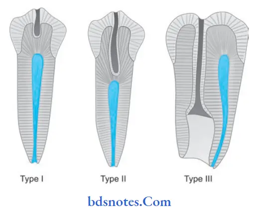
- Coronal dens invaginatus is of three types viz:
- Type I: Exhibit invagination which is confied to the crown.
- Type II: Extends below CEJ and ends in a blind sac that may or may not communicate with adjacent dental pulp.
- Type III: It extends through the root and perforates in apical or lateral radicular area without any immediate communication with the pulp.
“Common challenges in diagnosing dens invaginatus effectively: FAQs provided”
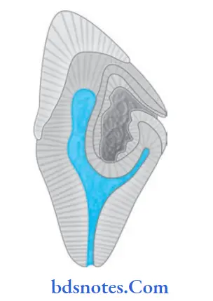
- Occasionally invagination can be rather large and resemble a tooth inside a tooth that’s why it is known as dens in dente.
- In some of the other cases invagination can be dilated and disturb the formation of tooth which lead to anomalous development of tooth known as dilated odontome.
- Roentgenographically, it is recognized as a pear shaped invagination of enamel and dentin with a narrow constriction at the opening on the surface of the tooth and closely approximating the pulp in its depth.
“Steps to explain causes of dens invaginatus: Genetic vs developmental factors: Q&A guide”
Radicular Dens Invaginatus
tooth within a tooth
- This condition is rare.
- This condition arises secondary to proliferation of
Hertwig’s epithelial root sheath, with formation of strip of enamel that extends along surface of root. - Roentgenographically, affected tooth shows enlargement of root. On close examination, there is presence of dilated invagination which is lined by the enamel with opening of invagination situated along lateral aspect of root.
“Role of enamel organ invagination in causing dens invaginatus: Questions answered”
“Early warning signs of issues addressed by understanding dens invaginatus pathogenesis: Common questions”
Treatment
- Type I invagination, opening should be restored after the eruption to prevent development of caries and pulpal inflammation.
- In large invagination content of lumen and carious dentine is removed and then calcium hydroxide base may be placed.
- Type III invaginations require endodontic therapy.
Leave a Reply