Deciduous Upper Second Molar: Buccal, Lingual, And Occlusal Anatomy
Describe in distal about Primary Maxillary Second Molar.
Answer:
Primary Maxillary Second Molar Buccal aspect:
- It shows two well-defined buccal cusps with a buccal developmental groove between them.
- The crown is narrow at the cervix.
- The roots appear slender and longer.
- The point of bifurcation between the buccal roots is close to the cervical line.
- The two buccal cusps are nearly equal in size.
Primary Maxillary Second Molar Lingual aspect:
- It shows three cusps.
- Mesiolingual cusp – large.
- Distolingual – well-developed.
- Supplemental cusp=apical to mesiolingual cusp called tubercle of carabelli.
- It is poorly developed.
- It acts as a buttress to the bulk of the mesiolingual cusp.
- If it is missing, some traces of developmental lines or “dimples” remain.
- A well-defined development groove separates the mesiolingual cusp from the distolingual cusp.
- It connects with the developmental groove.
- All three roots are visible.
- The lingual root is large and thick.
Primary Maxillary Second Molar Mesial aspect:
- The mesiolingual cusp appears large.
- The mesiobuccal cusp is short and sharp.
- Little curvature to the cervical line is seen.
- The mesiobuccal root is broad and flat.
- The lingual root appears long and slender.
- The point of bifurcation between the mesiobuccal root and the lingual root is 2-3 mm apical to the cervical line of the crown.
- The mesiolingual cusp is below the bifurcation.
- Cervical curvature is lingual.
Primary Maxillary Second Molar Distal aspect:
- A lingual outline creates a smooth, rounded line.
- Distobuccal and distolingual cusp are of the same length.
- The cervical line is straight.
- All three roots are seen.
- The distobuccal root is shorter and narrower.
- The point of bifurcation between the distobuccal root and lingual root is more apical.
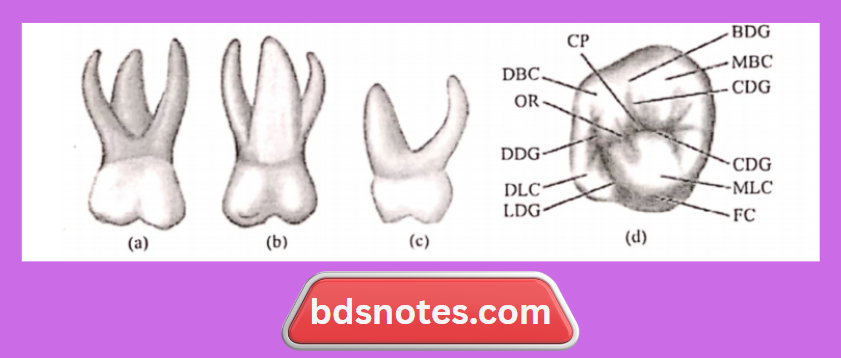
Primary Maxillary Second Molar Occlusal aspect:
- It is somewhat rhomboidal.
- It shows 4 cusps.
- Mesiobuccal.
- Distobuccal
- Mesiolingual
- Distolingual
- Supplemental cusps.
- The buccal surface is flat with a developmental groove.
- Occlusal surface has.
- A central fossa with a central pit.
- A well-defined mesial triangular fossa is distal to the mesial marginal ridge.
- The oblique ridge connects the mesiolingual and distobuccal cusp.
- Central groove connecting mesial triangular fossa with the central fossa.
- Buccal developmental groove.
- Extends buccally from the central pit.
- Separates triangular ridges.
- Lingual developmental groove.
- Separates mesiolingual and distolingual cusps.
- Supplemental grooves.
- Radiates from developmental grooves.
- Distal fossa- distal to oblique ridge.
- Distal triangular fossa.
- Mesial to the distal marginal ridge.
Leave a Reply