Cranial Cavity Question And Answers
Enumerate dural venous sinus. Describe the cavernous sinus in detail. (or)
Classify dural venous sinuses. Describe in detail its position, contents, tributaries & applied aspect of cavernous sinus. (or)
Describe the relation, tributaries, position, contents & connections of cavernous sinus. Add a note on applied anatomy. (or) Cavernous sinus, its relations & tributaries
Answer:
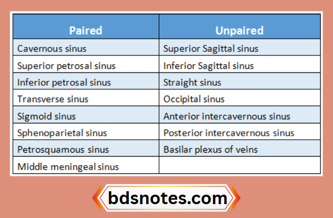
Dural Venous Sinuses:
- These are venous spaces whose walls are formed by duramater
- There are 23 venous sinuses- 8 paired & 7 unpaired
“Understanding the cranial cavity through FAQs: Structure, functions, and uses explained”

“Importance of studying the cranial cavity for medical students: Questions explained”
Read And Learn More: BDS Previous Examination Question And Answers
Cavernous Sinus:
Position:
It is situated in the middle cranial fossa on either side of the body of sphenoid bone
Boundaries:
- Floor- endosteal dura mater
- Lateral wall, roof & medial wall- meningeal dura mater
Extend:
- Anteriorly- upto medial end of superior orbital fissure
- Posteriorly- upto apex of petrous temporal bone
“Common challenges in mastering cranial cavity notes effectively: FAQs provided”
“Factors influencing success with cranial cavity studies: Q&A”
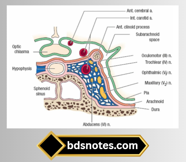
Relations:
1. Structures outside the sinus:
- Superiorly
- Optic tract
- Optic chiasma
- Olfactory tract
- Internal carotid artery
- Anterior perforated substance
- Inferiorly
- Foramen lacerum
- Junction between the body & greater wing of sphenoid
- Medially
- Hypophysis cerebri
- Sphenoidal air sinus
- Laterally
- Temporal bone with uncus
- Anteriorly
- Superior orbital fissure
- Apex of the orbit
- Posteriorly
- Apex of petrous temporal bone
- Crus cerebri of the midbrain
“Steps to explain the structure of the cranial cavity: Skull bones vs meninges: Q&A guide”
“Role of skull bones in forming the cranial cavity: Questions answered”
2. Structures in the lateral wall of the sinus:

3. Structures passing through the center of the sinus:
- Internal carotid artery with venous & sympathetic plexus
- Abducent nerve
Tributaries:
1. From the orbit
- Superior ophthalmic vein
- Branch of inferior ophthalmic vein
- Central vein of the retina
“Early warning signs of gaps in understanding structural principles: Common questions”
2. From the brain
- Superficial middle cerebral vein
- Inferior cerebral veins from the temporal lobe
3. From the meninges
- Sphenoparietal sinus
- Frontal trunk of the middle meningeal vein
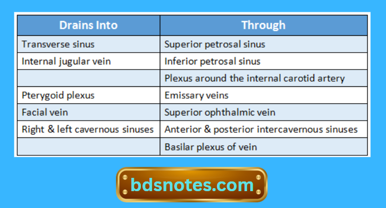
Communications:
“Asymptomatic vs symptomatic effects of ignoring cranial cavity basics: Q&A”
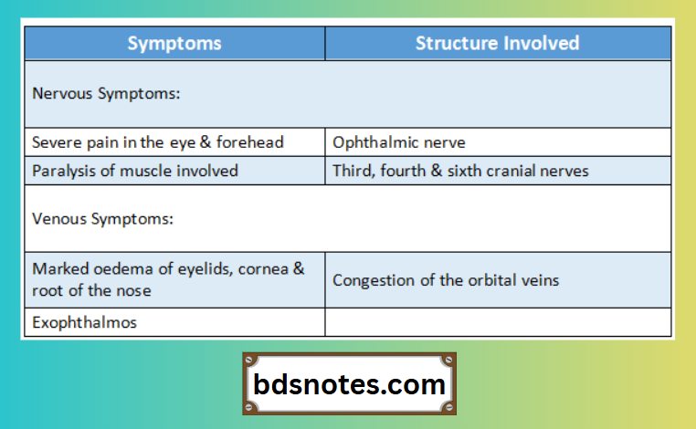
Applied Anatomy:
- Thrombosis of the cavernous sinus may be caused by sepsis in the dangerous area of the face, in nasal cavities & in the paranasal air sinuses
- The following symptoms occur.
“Differential applications of physical exams vs laboratory tests: Questions answered”
Leave a Reply