Chapter 2 General Characteristics Of Microbes Question And Answers
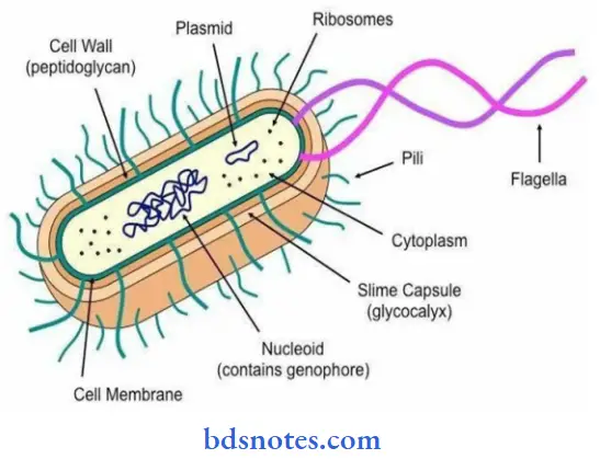
Question 1. Draw a neat labelled diagram of a Bacterial cell.
Answer:
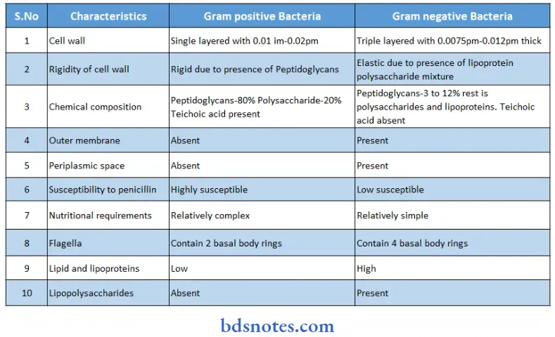
Question 2. Differentiate between Gram-positive bacteria and Gram-negative bacteria
Answer:
“Understanding general characteristics of microbes through FAQs: Q&A explained”
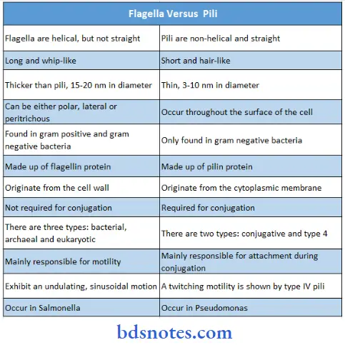
Question 3. Differentiate between Flagella and Fimbriae.
Answer:
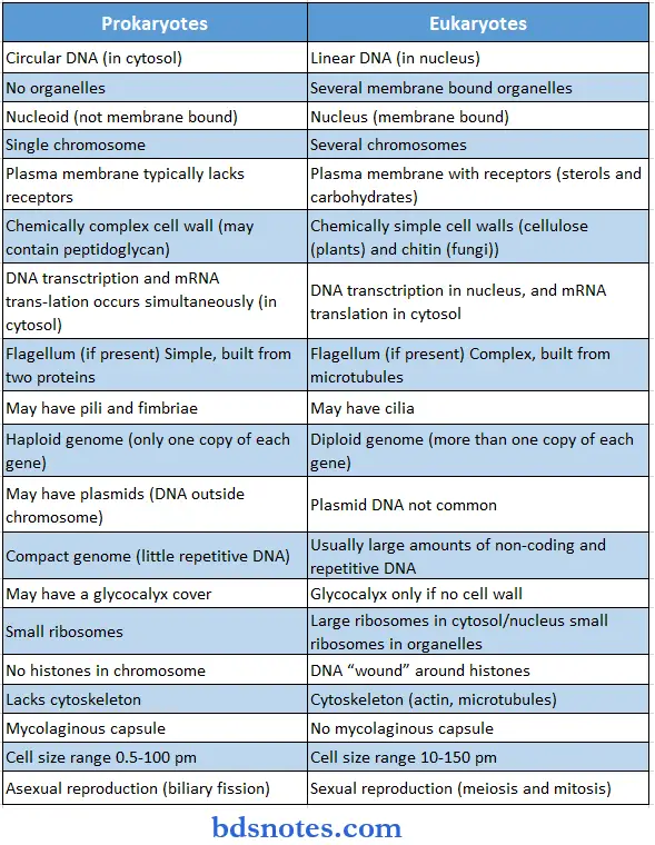
Question 4. Differentiate between Prokaryotic and eukaryotic cell.
Answer:
“Importance of studying microbes for BSc Nursing students: Questions explained”
Prokaryotic And Eukaryotic Cell Example:
- Fungi, protozoa, plants, animals
- Eubacteria, All bacteria, and blue-green algae
Question 5. Bacterial Growth curve.
Answer:
Bacterial Growth Curve:
- If a suitable liquid medium is inoculated with bacterium and incubated, its growth follows a definitive course.
- Small samples are taken at regular intervals after inoculation and plotted in relation to time. A plotting of the data will yield a characteristic growth curve.
- The changes of slope on such a graph indicate the transition from one phase of development to another.
Phases of Bacterial Growth Curve: The bacterial growth curve can be divided into four major phases:
- Lag phase
- Exponential or log (logarithmic) phase
- Stationary phase, and
- Decline phase.
These phases reflect the physiologic state of the organisms in the culture at that particular time.
- Lag Phase: After inoculation of the culture medium, multiplication usually does not begin immediately.
- The period between inoculation and beginning of multiplication is known as lag phase.
- During this period the organisms adapt to the new environment, during which necessary enzymes and intermediate metabolites are built up in adequate quantities for multiplication to proceed.
- There is an increase in the size of the cells but there is no appreciable increase in numbers.
- Log (Logarithmic) or Exponential Phase: The cell division starts and their numbers exponentially or by geometric progression with time. If the logarithm of the viable count is plotted against time, a straight line is obtained.
- Stationary Phase: After the log phase, bacterial growth ceases almost completely due to exhaustion of nutrients and accumulation of toxic products. The number of progeny cells formed is just enough to replace the number of cells that die. The number of viable cells remains stationary as there is almost a balance between the dying cells and the new formed cells.
- The phase of Decline: Alter a period of stationary phase, the bacterial population decreases due to the death of cells. The decline phase starts due to the exclusion of nutrients, accumulation of toxic produce, and autolytic enzymes. There is decline in viable count and not in total count.
Read And Learn More: Bsc Nursing 1st Year Microbiology Previous year Question and Answers

“Common challenges in understanding characteristics of microbes effectively: FAQs provided”
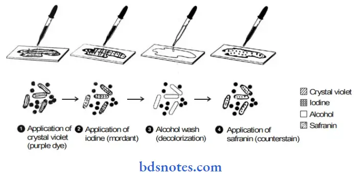
Question 6. Gram Staining.
Answer:
The technique was developed by a Danish physician. Dr hanes Cristain Gram 1884. This is a useful differential staining procedure in bacteriology which besides determining gross morphology differentiates
Bacteria into two major distinct groups:
- Gram Positive
- Gram Negative
The technique involves six basic steps:
- Smear preparation
- Heat fixing of smear
- Staining with a crystal violet (Primary staining)
- Use of iodine.
- Treatment with acetone alcohol mixture (Decolourizing agent).
- Use of safarin (Colour stain).
Gram Staining Principale: The peculiar response toward the staining is related to physical and chemical differences in the cell wall of the two groups of bacteria.
- In gram-negative bacteria the cell wall is thin and multilayered containing high lipid contents that are readily dissolved by alcohol, resulting in pore formation in the cell wall facilitating the leakage of the crystal violet iodine complex of resulting in discoloration of gram-negative
bacteria which takes safarin and appears red. - On the other hand. Cell walls of gram-positive bacteria are thick, composed mainly of proteins and crossed-linked mucopeptides on the application of a decolorizing agent, dehydration results in closure of pores of cell wall thereby retaining the CV-1 complex and appearing blue or Purple.
“Why is early learning of microbial characteristics critical for nursing practice? Answered”
Gram Staining Reagents Used:
- Crystal violet
- Gram iodine solution
- Ethyl alcohol (15%) or Alcohol acetone (1:1) solution.
- Safranin Solution
Gram Staining Procedure:
- Make smear of a given culture on a clear glass slide.
- Air dry the smear and heat fix it.
- Cover the smear completely with crystal violet stain and leave the stain on the slide for one minute.
- Wash the slide gently in distilled water or tap water
- Flood the spray with gram iodine solution for 1 minute.
- Wash with tap water gently and drain carefully.
- Wash the slide gently under running water for 30 min.
- Now counter-stain with Safari and wait for 30 min
- Wash again and flot dry with bloting paper or simply air dry the slide and observe under oil. Immersion objective.
Gram Staining Result:
- Bacteria that appear blue / violet/purple are assigned as gram-positive.
- While those appearning red/pink are assigned as gram negative.
Question 7. Acid-fast staining.
Answer:
- The technique was developed by Paul Ehrlich. and was modified later by Ziehl Nelson.
- This is a differential staining used to identify mainly the members of mycobacterium, especially these organisms are difficult to stain by ordinary staining method due to the presence of high lipid content in their cell walls.
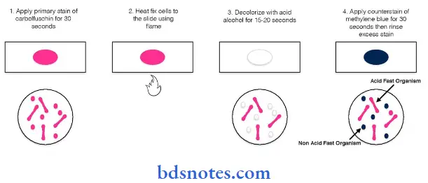
Bacteria are Classified as:
- Acid-fast: They retain primary stain after the application of strong the application of strong acid and appear red
- Non-acid fast: if they not retain the primary stain and are counter-stained by methylene blue.
Acid Fast Staining Reagent:
- Carbol fusion solution.
- Acid alchol solution (20 – 25 % v/v in water)
- Methylene blue counter stain (0.3 w/v aqueous)
Acid Fast Staining Procedure:
- Prepare a smear of purulent portion of the specimen on a clean glass slide
- Air dry and heat fix the smear
- Flood the smear with freshly filtered carbol Fuchisin. Heat gently until steam rises.
- Cool and wash the stain of the slide with H2O
- Cover the slide with an acid alcohol solution for 3 min. Wash with running tap water and drain Finally, wash it.
- Cover the slide with methylene blue stain and leave it for 2 min.
- Wash with tap water, blot dry or air dry the slide, and observe under an oil immersion object.
Acid Fast Staining Result: Acid-fast organisms will appear bright red on a blue background. While as nonacid fast organisms will appear dark blue in colour.
“Steps to explain the structure of microbes: Prokaryotes vs eukaryotes: Q&A guide”
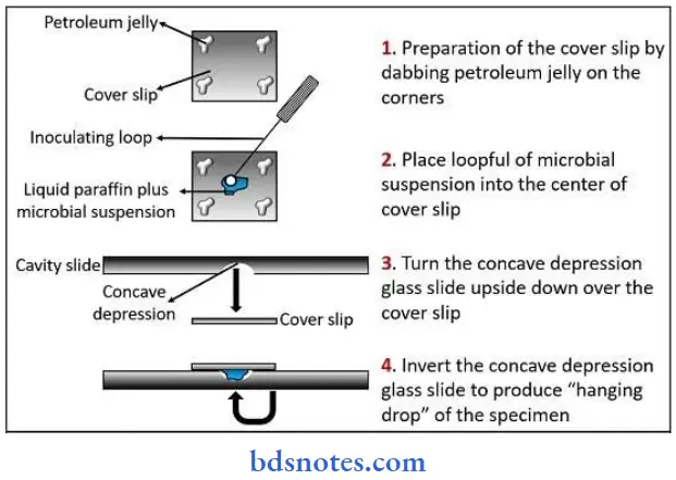
Question 8. Hanging drop method.
Answer:
Hanging drop preparation is a special type of wet mount (in which a drop of medium containing the organisms is placed on a microscope slide), often is used in dark illumination to observe the motility of bacteria.
Hanging Drop Method Preparation
- In this method, a drop of culture is placed on a coverslip that is encircled with petroleum jelly (or any other sticky material).
- The coverslip and drop are then inverted over the well of a depression slide. The drop hangs from the coverslip, and the petroleum jelly forms a seal that prevents evaporation. This preparation gives good views of microbial motility.
Hanging Drop Method Materials Required
- Glass slides (glass slide with depression) or normal glass slide with adhesive or paraffin ring
- Paraffin wax
- Loop
- Coverslip
- Microscope
- Bunsen burner
- Young broth culture of motile bacteria (for example, Proteus mirabilis)
Hanging Drop Method Procedure
- Take a clean glass slide and apply a paraffin ring, and adhesive tape ring to make circular concavity. (This step is not needed if a glass slide with depression is available).
- Hold a clean coverslip by its edges and carefully dab Vaseline on its corners using a toothpick.
- Place a loopful of the broth culture to be tested in the center of the prepared coverslip.
- Turn the prepared glass slide or concavity slide upside down (concavity down) over the drop on the coverslip so that Vaseline seals the coverslip to the slide around the concavity.
- Turn the slide over so the coverslip is on top and the drop can be observed hanging from the coverslip over the concavity.
- Place the preparation in the microscope slide holder and align it using the naked eye so an edge of the drop is under the low power objectives.
- Turn the objective to its lowest position using the coarse adjustment and Close The Diaphragm.
- Look through the eyepiece and raise the objective slowly using the coarse adjustment knob until the edge of the drop is observed as an irregular line crossing the field.
- Move the slide to make that line (the edge of the drop) pass through the centre of the field.
- Without raising or lowering the tube, swing the high dry objective into position (Be sure the high dry objective is clean).
- Observe the slide through the eyepiece and adjust the fine adjustment until the edge of the drop can be seen as a thick, usually dark line.
- Focus the edge of the drop carefully and look at each side of that line for very small objects that are bacteria. The cells will look either like dark or slightly greenish, very small rods or spheres. Remember the high dry objective magnifies a little less than half as much as the oil immersion objective.
- Adjust the light using the diaphragm lever to maximize the visibility of the cells.
- Observe the cells noting their morphology and grouping and determine whether true motility can be observed.
- Brownian movement should be visible on slides of all the organisms, but there should also show true motility.
- Wash the depression slide and after soaking in lysol buckets or discard the prepared glass slide.
“Role of microbial morphology in identification: Questions answered”
Hanging drop slides are useful in observing the general shape of living bacteria and the arrangement of bacterial cells when they associate together. Organisms are observed in a drop that is suspended under a cover glass in a concave depression slide.
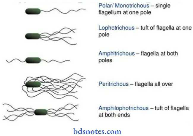
Question 9. Bacterial flagella.
Answer:
Bacterial Flagella: Motile bacteria, except spirochetes, possess one or more unbranched, long, sinuous filaments called flagella, which are the organs of locomotion.
Bacterial Flagella Structure: They are long, hollow, helical filaments, usually several times the length of the cell. They are 3-20 µm long and are of uniform diameter (0.01-0.013 µm) and terminate in a square tip. Flagella can be found on both gram-positive and gram-negative bacilli.

Flagella Parts and Composition: Each flagellum consists of three parts
- Filament
- Hook
- Basal body.
“How do size and shape define microbial behavior? FAQ explained”
- Filament: The filament is the longest and most Obvious portion which extends from the cell surface to the tip.
- Hook: The hook is a short, curved segment that links the filament to its basal body and functions as the universal joint between the basal body and the filament.
- Basal body: The basal body is embedded in the cell (cytoplasmic membrane). In gram-negative bacteria, the basal body has four rings connected to a central rod (L, P, S, and M).
Gram-positive bacteria have only two basal body rings, an inner ring connected to the cytoplasmic membrane and an outer one probably attached to peptidoglycan.
Flagella Arrangement Or Types:
- The number and location of flagella are distinctive for each genus. There are four types of flagella arrangement:
- Monotrichous—Single polar flagellum (example, Cholera vibrio).
- Amphitrichous—Single flagellum at both ends (example, Alcaligenes faecalis). Types of flagella arrangement
- Lophotrichous—Tuft of flagella at one or both ends (example, spirilla).
- Peritrichous—Flagella surrounding the cell (for example, Typhoid bacilli).
- Flagella are about 0.02 µm in thickness and hence beyond the resolution limit of the light microscope.
“Early warning signs of gaps in understanding microbial basics: Common questions”
Question 10. Bacterial Spore
Answer:
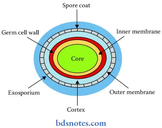
Bacterial Spore
- A number of gram-positive bacteria, such as those of the genera Clostridium and Bacillus can form a special resistant dormant structure called an endospore or, simply, spores.
- Endospores develop when essential nutrients are depleted. In sporulation, each vegetative cell forms only one spore, and in subsequent germination, each spore gives rise to a single vegetative cell.
- Sporulation in bacteria, therefore, is not a method of reproduction but of preservation.
Bacterial Sporulation: Spore formation, sporogenesis or sporulation normally commences when growth ceases due to lack of nutrients, depletion of the nitrogen or carbon source (or both) being the most significant factor. New antigens appear on sporulation that are not found in the vegetative cell.
Bacterial Spore Stages: It is a complex process and may be divided into several stages.
- Bacteria Spore septum: In the first observable stage of sporulation, a newly replicated bacterial chromosome and a small portion of cytoplasm are isolated by an ingrowth of the plasma membrane called a spore septum.
- Forespore: The spore septum becomes a double-layered membrane that surrounds the chromosome and cytoplasm. Structure, entirely enclosed within the original cell, is called a forespore.
- Spore coat: The forespore is subsequently completely encircled by dividing septum as a double-layered membrane. The two spore membranes now engage in active synthesis of various layers of the spore. The inner layer becomes the inner membrane. Between the two layers is laid spore cortex and outer layer is transformed into a spore coat which consists of several layers. In some species from outer layer also develops exosporium which bears ridges and folds.
- Free endospore: Finally exosporium disintegrates and the endospore is freed.

“Asymptomatic vs symptomatic effects of ignoring microbial characteristics: Q&A”
Question 11. Explain in detail about morphology of bacteria and structural components of bacterial cell along with their function.
Answer:
- Bacteria are very small in size. The unit of measurement in bacteriology is the micron or micrometer (mm).
- Bacteria of medical importance generally measure 0.2-1.5 µm in diameter and about 3-5 µm in length. To see bacteria, a light microscope must be used.
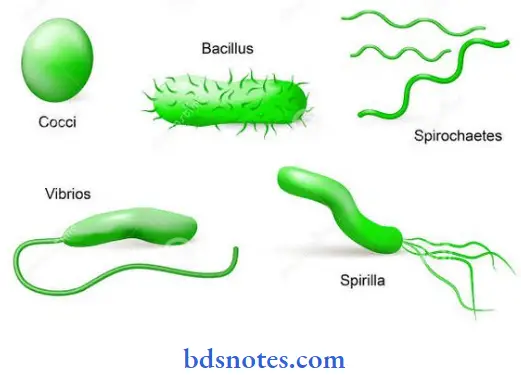
Shape of Bacteria Depending on their shape, bacteria are classified into several varieties:
- Cocci: Cocci (from kokkos meaning berry) are spherical, or nearly spherical.
- Bacilli: Bacilli (from bacillus meaning rod) are relatively straight, rod-shaped (cylindrical) cells. In some of the bacilli, the length of the cells may be equal to width. Such bacillary forms are known as lactobacilli and have to be carefully differentiated from cocci.
- Vibrios: Vibrios are curved or comma-shaped rods and derive their name from their characteristic vibratory motility.
- Spirilla: Spirillas are rigid spiral or helical forms.
- Spirochetes: Spirochetes (from spiral meaning coil and chaite meaning hair) are flexuous spiral forms.
- Mycoplasma: Mycoplasma are cell wall deficient bacteria and hence do not possess a stable morphology. They occur as round or oval bodies and interlacing filaments.
“Can targeted interventions improve outcomes using microbial knowledge? FAQs provided”
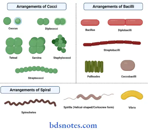
Cocci Arrangement
- Diplococci: Cocci may be arranged in pairs (diplococci) when cocci divide and remain together.
- Long chains: Long chains (Streptococcus, Enterococcus, and Lactococcus) when cells adhere after repeated divisions in one plane.
- Grape-like clusters: Grape-like clusters (staphylococci) when cocci divide in random planes.
- Tetrads: Square groups of four cells (tetrads) when cocci divide in two planes as in members of the genus Micrococcus.
- Cubical packets: Cubical packets of eight of cells (genus Sarcina) when cocci divide in three planes.
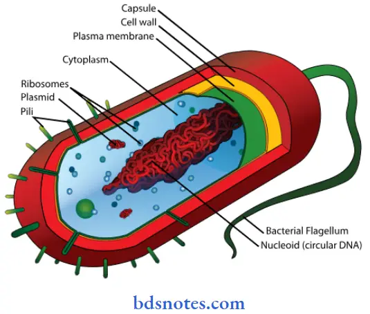
Structural components of bacterial cell: Bacterial Cell Components can be divided into
The outer layer or cell envelope consists of two components:
- Cell wall.
- Cytoplasmic or plasma membrane—beneath cell wall.
Cellular appendages—Besides these essential components, some bacteria may possess additional structures such as capsules, fimbriae, and flagella.
Capsule: Some bacteria produce a protective gelatinous covering layer called a capsule outside the cell wall. If the capsule is too thin to be seen with light microscope (<0.2 µm) it is called a microcapsule.
Loose slime: Soluble, large-molecular, amorphous, viscid colloidal material may be dispersed by the bacterium into the environment as loose slime.
Flagella: Some bacteria carry external filamentous appendages protruding from the cell wall flagella, which are organs of locomotion fimbriae, which appear to be organs of adhesion and pili, which are involved in the transfer of genetic material.
The Outer Layer or Cell Envelope
1. Cell Wall: The cell wall is the layer that lies just outside the plasma membrane. It is 10-25 nm thick, strong, and relatively rigid, though with some elasticity, and openly porous, being freely permeable to solute molecules smaller than 10 kDa in mass and 1 nm in diameter.
“Early warning signs of complications from ignoring microbial traits: Common questions”
“Asymptomatic vs symptomatic effects of outdated microbial practices: Answered”
Functions of the cell wall:
- To impart shape and rigidity to the cell.
- It supports the weak cytoplasmic membrane against the high internal osmotic pressure of the protoplasm (ranges from 5 and 25 atm).
- Maintains the characteristic shape of the bacterium.
- It takes part in cell division.
- Also functions in interactions (for example, adhesion) with other bacteria and with mammalian cells.
- Provide specific protein and carbohydrate receptors for the attachment of some bacterial viruses.
Chemical Structure of Bacterial Cell Wall: Chemically the cell wall is composed of mucopeptide (peptidoglycan or murein) scaffolding formed by N-acetyl glucosamine and N-acetyl muramic acid molecules alternating in chains, which are crosslinked by peptide bonds. Peptidoglycan consists of three parts
- A backbone— composed of alternating N-acetylglucosamine and N-acetylmuramic acid.
- A set of identical tetrapeptide side chains attached to N-acetylmuramic acid.
- A set of identical pentapeptide cross-bridges
“Steps to apply microbial characteristics in clinical practice: Identification vs diagnosis: Q&A guide”
Question 12. Define culture media and classify them on the basis of various methods in detail.
Answer:
Culture Medium Introduction: A nutrient material prepared for the growth of microorganisms in a laboratory is called a culture medium.
Classification Of Culture Media: Media have been classified into many ways
1. Phases Of Growth Media: Growth media are used in either of the two phases.
- Liquid (Broth) Media: In broth media, nutrients are dissolved in water, and bacterial growth is indicated by a change in broth’s appearance from clear to turbid (i.e. cloudy).
- Solid (Agar) Media: Solid media are made by adding a solidifying agent to the nutrients and water. Agarose is the most common solidifying agent. The Petri dish containing the agar is referred to as agar.
- Semisolid Media: For special purposes where agar is added to media in concentrations that are too low to solidify them.
2. Based On Nutritional Factors
- Simple media (Basal media): Simple media are those which contain only basic nutrients required for the growth of ordinary organisms, and are used as a general-purpose media, for example, peptone water, nutrient broth, and nutrient agar.
- Complex media: Media that contain some ingredients of unknown chemical composition are called complex media. One common ingredient is peptone. Extracts, which are the water-soluble components of a substance, are also used.
- Synthetic Or Chemically Defined Media: They are prepared exclusively from pure chemical substances and their exact composition is known.
“How does microbial diversity impact healthcare practices? FAQ explained”
3. Special Media
- Enriched Media: These are prepared to meet the nutritional requirements of more exacting bacteria by the addition of substances such as blood, serum, or egg to a basal medium.
- Examples Of Enriched Media
- Blood agar
- Chocolate agar
- Examples Of Enriched Media
- Selective Media: When a substance is added to a solid medium that inhibits the growth of unwanted bacteria but favors the growth of wanted bacteria, it is known as selective media. These media are used to isolate particular bacteria from specimens where mixed bacterial flora is expected.
- Examples of selective media:
- Deoxycholate citrate agar (DCA): The addition of deoxycholate acts as a selective agent for dysentery bacilli (isolation of Shigellae).
- Lowenstein-Jensen medium: This medium is used for Mycobacterium tuberculosis.
- Bile salt agar (BSA): Bile salt is a selective agent. It farceurs the growth of only Vibrio cholerae and inhibits the growth of intestinal organisms.
- Examples of selective media:
- Indicator Media: These media contain an indicator that changes colour when a bacterium grows in them.
- Indicator Media Examples:
- Wilson and Blair medium:
- MacConkey agar
- Indicator Media Examples:
- Differential Media: A medium, that has substances incorporated in it, enabling it to bring out differing characteristics of bacteria and thus helping to distinguish between them, is called a differential medium.
- Differential Media Example:
- MacConkey agar: MacConkey agar is both differential and selective.
- Differential Media Example:
- Sugar Media: For the identification of most of the organisms, sugar fermentation reactions are carried out. Carbohydrate fermentation is used ‘for the characterization and identification of bacteria, particularly important in the study of Gram-negative bacilli. Sugar media are used to test fermentation.
- Sugar used for sugar media: The term ‘sugar’ in microbiology denotes any fermentable
substance. Glucose, lactose, sucrose, and mannitol are routinely employed for fermentation tests.
- Sugar used for sugar media: The term ‘sugar’ in microbiology denotes any fermentable
- Transport Media: A transport medium is a holding medium designed to preserve the viability of microorganisms in the specimen but not allow multiplication.
- Transport Media Examples
- Stuart’s transport medium and Amines transport medium for gonococci.
- Buffered glycerol saline for enteric bacilli.
- Transport Media Examples
“Role of microbial traits in disease prevention: Questions answered”
Question 13. Define and Draw a neat labelled diagram of bacterial spore.
Answer:
- Bacterial spores serve largely as a resting, or dormant, stage in the bacterial life cycle, helping to preserve the bacterium through periods of unfavorable conditions.
- Spore production is particularly common among Bacillus and Clostridium bacteria, several species of which are disease-causing.
Leave a Reply