Blood Disorders Question And Answers
Question 1. What is anemia? Classify and describe clinical manifestation, laboratory diagnosis and treatment planning of iron deficiency anemia.
Or
Define and classify anemia. Discuss clinical features, laboratory investigations, oral manifestations and management of iron deficiency anemia.
Answer. Anemia is defined as an abnormal reduction in number of circulating RBCs, the quantity of hemoglobin and volume of packed red cell in given unit of blood.
Classification of Anemia
- Anemia due to increased blood loss:
- Acute posthemorrhagic anemia
- Chronic posthemorrhagic anemia
- Anemia due to impaired red cell function:
- Cytoplasmic maturation defect:
- Deficient heme synthesis, i.e. iron deficiency anemia.
- Defective globin synthesis, i.e. thalassemic syndrome
- Nuclear maturation defect, i.e. vitamin B12 or folic acid deficiency, e.g. megaloblastic anemia
- Defect in stem cell proliferation and differentiation
- Aplastic anemia
- Pure red cell aplasia
- Anemia of chronic disorders
- Bone marrow infiltration
- Congenital anemia
- Cytoplasmic maturation defect:
- Hemolytic anemia
- Acquired or extracorpuscular
- Immunohemolytic anemia
- Autoimmune hemolytic anemia
- Warm antibody autoimmune hemolytic anemia
- Cold antibody autoimmune hemolytic anemia
- Drug-induced immunohemolytic anemia.
- Isoimmune hemolytic anemia.
- Autoimmune hemolytic anemia
- Mechanical trauma: Microangiopathic hemolytic anemia
- Direct toxic effect: Malaria, bacteria, infection and other agents.
- Acquired red cell membrane abnormalities: Paroxysmal nocturnal hemoglobinuria.
- Splenomegaly
- Hereditary or intracorpuscular
- Abnormalities of red cell membrane:
- Hereditary spherocytosis.
- Hereditary elliptocytosis.
- Hereditary stomatocytosis.
- Disorders of red cell interior:
- Red cell enzyme defect:
- Defects in HMP shunt: G6PD deficiency.
- Defects in glycolytic pathway: Pyruvate kinase deficiency.
- Disorders of hemoglobin:
- Structurally abnormal hemoglobin, sickle syndrome and other hemoglobinopathies.
- Reduced globin chain synthesis: Thalassemias.
- Red cell enzyme defect:
- Morphologic:
- Microcytic hypochromic
- Normocytic normochromic
- Macrocytic normochromic.
Read And Learn More: Oral Medicine Question And Answers
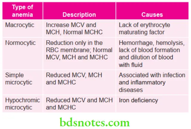
Classification of Iron Deficiency Anemia
Anemia Clinical Manifestations
- It occurs in women in 4th to 5th decades of life.
- Patient experiences, tiredness, headache, paresthesia and lack of concentration.
- Koilonychia: Nails become flattened, brittle and spoon shaped.
- Neuropathy: There may be pin and needle sensation in extremities.
- Dysphagia: Some patients develop pharyngeal mucosal thickening.
- Gastrointestinal symptoms: Liver and spleen may be palpable. There may be gartrointestinal bleeding. Menorrhagia is present.
- Knuckle pigmentation can be appreciated.
Anemia Oral Manifestations
- There is pallor of oral mucosa and gingiva.
- There is generalized atrophy of oral mucosa in tongue and buccal mucosa
- Tongue changes:
- There is redness, soreness or burning of tongue
- The filiform papilla over the anterior two-thirds of tongue is first to undergo atrophy
- In severe cases, fungiform papilla are affected leaving tongue smooth and waxy.
- Angular cheilitis: There is cracking and fissuring at the corners of mouth
- Softening of epithelium which leads to linear ulceration of skin, ascending upto and beyond the mucocutaneous junction. Bleeding from ulcerated tissues is appreciated
- Recurrent aphthous ulceration and candidal lesions can also occur in iron deficiency anemia
- Patient shows slow healing after oral surgical procedures.
Anemia Laboratory Diagnosis
- Anemia is microcytic and hypochromic and peripheral smear shows abnormal forms of RBCs.
- There is reduced hemoglobin level, as low as 4 g/100 mL.
- There is normal or slightly reduced red cell count. MCV, MCH, MCHC all are reduced.
Anemia Treatment Planning/Management
- Iron supplement: Almost all patients are treated with oral supplements of iron by giving ferrous fumarate or ferrous sulfate. It is given in dose of 300 mg TDS or QDS for period of 6 months.
- The parental route of administration is suitable for few patients who are unable to take iron by mouth or who are unable to absorb iron by mouth.
- Recommended single dose of iron sorbitol is 1.5 mg/kg of body weight given daily.
- Parenteral iron supplements should be given at dose of 15 mg/kg body weight till maximum of 750 mg on two occasion atleast 7 days apart.
Question 2. Write short note on iron deficiency anemia.
Answer. Iron deficiency anemia is caused by the imbalance between iron intake and loss or inadequate utilization.
Iron Deficiency Anemia Causes
- Inadequate intake of iron
- Malabsorption of iron due to hypochlorhydria and diarrhea
- Increased requirement of iron in a growing child and pregnancy
- Increased loss of iron due to injury, recurrent epistaxis and peptic ulcer
- Chronic blood loss such as menstrual and menopausal bleeding parturition
- Subtotal or complete gastrotomy.
Question 3. Write short note on clinical features (with emphasis on oral manifestation) and diagnosis of pernicious anemia.
Or
Write short note on clinical features and laboratory investigations of Pernicious Anemia.
Answer. The pernicious anemia is also called as primary anemia, Addison’s anemia or Biermer’s anemia.
The term pernicious anemia is given to the patients who have vitamin B12 deficiency which is secondary to intrinsic factor deficiency.
Pernicious Anemia Clinical Features
- It is rare before the age of 30 years and increase frequently with advancing age. Males are more commonly affected than females.
- There is a triad of symptoms, i.e. generalized weakness, sore painful tongue and numbness and tingling of extremities.
- Other features are fatigability, headache, dizziness, nausea, vomiting, diarrhea, with loss of appetite, shortness of breath, loss of weight, pallor and abdominal pain.
- In nervous system, sensory disturbances are present such as paresthesia of extremity, stiffness as well as difficulty in the walking, irritability and depression, drowsiness and incoordination of loss of vibratory sensation. Presence of tingling sensation in finger and toes which progresses to numbness.
- Presence of gastrointestinal symptoms such as constipation, diarrhea and epigastric discomfort.
Pernicious Anemia Oral Manifestations
- Presence of glossitis and patient complains of painful and burning lingual sensation.
- The tongue is inflamed and is beefy red in color within entirely or in patches scattered over dorsum and lateral border of tongue.
- There is atrophy of papillae of tongue which eventuates in smooth and bald tongue which is referred to as Hunter’s glossitis or Moller’s glossitis.
- The fiery red appearance of tongue may undergo remission but recurrent attacks are common.
- There is disturbance in taste sensations with intolerance to dentures and occasional dryness of mouth.
- Oral mucosa shows greenish yellow color at junction of hard and soft palate, when day light is used for illumination.
Pernicious Anemia Diagnosis
The diagnosis is based on the laboratory investigations, i.e.
- RBC count is decreased often to 3 or less per cubic mm
- Many of cells exhibits macrocytosis, while some exhibits poikilocytosis
- Decreased WBC count and MCH
- Bone marrow shows great number of immature red cells or megaloblasts with few normoblasts
- Schilling test detects the absence of intrinsic factor.
- In advanced cases, there is presence of polychromatophilic cells, stippled cells, nucleated cells, Howell-Jolly bodies and cobat’s ring.
The above tests confirm the diagnosis.
Question 4. Write short note on pernicious anemia.
Answer.
Pernicious Anemia Causes
- It occurs due to atrophy of gastric mucosa resulting in failure to secrete still unidentified intrinsic factor.
- It is suggested that it is an autoimmune disorder, because autoantibodies to gastric parietal cells are often found in patients.
Pernicious Anemia Management
- For first week daily subcutaneous administration of vitamin B12 is given. If reticulocytosis is seen, then administration of vitamin B12 twice weekly for around 4 to 5 weeks. As this is over 100 µg of vitamin B12 should be administered monthly either subcutaneous or intramuscular route.
- Patient who does not consume vitamin B12 rich foods such as eggs, meat or milk should be given supplemental vitamin B12. 100 to 200 µg should be given orally either in tablet or in syrup form.
- Physical activity should be done.
Question 5. Write short note on Aplastic Anemia
Answer. It is a rare disorder characterized by peripheral blood pancytopenia (anemia, leukopenia and thrombocytopenia associated with bone marrow suppression)
Aplastic Anemia Types
- Primary: It is the disease of unknown origin which occurs most frequently in young adults, develops more rapidly and terminates fatally.
- Secondary: It has known etiology which occurs at any age and present better prognosis.
- Etiology
- Drugs and chemicals: Drug-related aplastic anemia develop in three ways, i.e.
- Characterized by myelosuppressive effect of cancer chemotherapeutic agents.
- Some drug can cause marrow depression.
- The third type is idiosyncratic or hypersensitivity induced aplastic anemia.
- Common drugs are benzene derivatives, chloramphenicol, amidopyrins organic arsenicals, colloidal silver, bismuth, mercury, sulfonamides, penicillin and anti-cancer drugs.
- Infections: Patients with bacterial disease such as tuberculosis and viral infections like hepatitis and infection mononucleosis can cause pancytopenia.
- Radiation: Long-term continuous exposure, to small amounts of external radiation or to internally deposited radium or thorium has been followed by development of aplastic anemia.
- Other causes: Pregnancy and thymoma can also cause aplastic anemia.
Aplastic Anemia Clinical Features
- It can occur at any age, but is common in young adults.
- Patient may feel weakness after slight physical exertion and exhibit pallor of skin
- Breathlessness, headache, ankle edema are common
- Numbness and tingling of extremities with edema
- Anemia may be severe enough to cause anginal pain or congestive cardiac failure
- There may be bleeding from various sites such as skin, nose, vagina and gastrointestinal tract associated with fever due to infection.
Aplastic Anemia Oral Manifestations
- Oral mucosa is pallor.
- In some of the cases, spontaneous hemorrhage may occur from gingiva.
- Petechiae, often are present on the soft palate and in severe cases, there may be submucosal ecchymosis
- Large-ragged ulcers which are covered by gray or black necrotic membrane may be present, which results in generalized lack of resistance to infection and trauma.
Aplastic Anemia Diagnosis
- Clinical diagnosis: Features such as thrombocytopenia, deficiency of WBCs i.e. presence of infection, deficiency of erythrocytes i.e. presence of pallor provide clue to diagnosis.
- Laboratory diagnosis or investigations:
- Decreased RBC count as low as 1 million cells/mm3.
- Count of WBCs is 2000 cells/mm3
- Platelet count falls below 20,000 cells/mm3
- Decrease in absolute reticulocyte count.
- Prolonged bleeding time and normal clotting time.
- Normocytic anemia along with some degree of macrocytosis.
- Bone marrow is fatty with few developing cells.
Aplastic Anemia Management
- Withdrawal of etiologic agent
- Supportive therapy should be in form of antibiotics and transfusion
- Stimulation of hemopoiesis by androgens
- Bone marrow transplantation can be useful in some cases
- Antifibrinolytic agents: Gingival bleeding can be reduced by using systemic antifibrinolytic agents such as aminocaproic acid or tranexamic acid and local treatment.
Question 6. Write short note on oral manifestations of leukemia.
Answer. Oral manifestations of leukemia:
Acute Leukemia
The submental, cervical, pre and postauricular lymph nodes may be enlarged and tender.
Leukemia Symptoms
- Paresthesia of lower lip and chin is present.
- There may be toothache due to leukemic cell infiltrationof dental pulp.
Leukemia Signs
- The oral mucus membrane shows pallor, ulceration of necrosis, petechial, ecchymosis and bleeding tendency.
- There may be massive necrosis of lingual mucosa with slough-ing, gingiva shows hypertrophy and cyanotic discoloration.
- The gingiva appears boggy, edematous and deep red, bleeds easily due to ulceration of sulcus epithelium and necrosis of underlying tissue.
- Mobility of permanent tooth is present.
- Oral infections (candidal, viral and bacterial) are serious and potentially fatal complication in leukemic patient.
Chronic Leukemia
- Gingiva: Most common oral finding is hypertrophy of gingiva. There may be ulceration with necrosis and gangrenous degeneration.
- Tongue: The tongue is swollen and dark.
- Lymph nodes: There is regional lymphadenopathy.
- Teeth: Rapid loosening of teeth due to necrosis of PDL. Destruction of alveolar bone occurs.
Question 7. Describe hemorrhagic disorder affecting oral cavity.
Answer. The hemorrhagic disorders are described as follows:
Hemophilia A
It is a hereditary disorder of blood coagulation characterized by excessive hemorrhage due to prolonged coagulation time. It is caused due to deficiency of factor VIII or antihemophilic factor.
Males are clinically affected and females are carriers of trait.
Hemorrhagic Disorder Oral Manifestations
- Traumatic injury of the oral cavity.
- The anatomic sites involved are frenum of lip and tongue
- Prolong bleeding after tooth extraction.
- Hematoma of floor of mouth may occur and blood may spread via facial spaces and produce hematoma of larynx, with respiratory embarrassment.
- Physiological processes of tooth eruption and exfoliation is associated with severe and prolonged hemorrhage.
Hemorrhagic Disorder Management
- Main aim is to raise factor VIII levels which arrest bleeding.
- Replacement therapy: Various forms of replacement therapy are like plasma, cryoprecipitate and factor VIII concentrates.
- Factor VIII concentrates: The dose and duration of administration of factor VIII concentrates are planned, taking into consideration in the site and type of bleeding.
- Intraligamentary injection in hemophilia does not cause hemorrhage, even if replacement therapy has not given prior to injection.
- To reduce complications caused by cryoprecipitate or plasma various drugs are used instead of factor VIII products, before dental extraction, e.g. a synthetic analog of 8-D-arginine and in combination with tranexamic acid and epsilon aminocaproic acid can be given.
Christmas Disease or Hemophilia B
It occurs due to hereditary deficiency of factor IX. It is transmitted as X-linked recessive character through chromosome.
Dental Management of Hemophilia A and Hemophilia B
- Anesthesia: It is given as intrapulpal, periodontal and papillary injection. Sedation with diazepam or nitrous oxide analgesia can be given.
- Endodontic therapy: During endodontic therapy, care is taken not to exceed beyond the apex of tooth. Hemorrhage into the canal can be controlled by 1:1000 aqueous epinephrine on an endodontic paper point.
- Restorative: It can be carried out in the hemophilic patient, but the rubber dam must be used to prevent trauma to gingiva and other soft tissues. If rubber dam is not used epinephrine impregnated hemostatic cord is placed in gingival sulcus before preparation of crown or inlay margins.
- Prosthodontic therapy: Complete dentures are well tolerated in hemophilic patients. Partial dentures are also well tolerated, as long as patient maintain oral hygiene because clasp can trap food debris, resulting in gingivitis and subsequent hemorrhage.
- Periodontal therapy: Conservative periodontal therapy is more desirable.
- Oral surgical procedure: Local hemostatic agents and techniques include pressure surgical pack, oxidize cellulose saturated with bovine thrombin solution, vasoconstrictions, sutures, topical thrombin and use of absorbable hemostatic after packing the tooth socket is prevented by mechanical splint to prevent disturbance to clot. Postoperative use of anti-fibrinolytic agent and adherence to soft diet are further advocated to support clot maintenance.
- Aspirin and other NSAID
- s are avoided in patients with bleeding disorders owing to inhibit their platelet function, IM injection is not given due to risk of hematoma formation.
- Electrosurgery is not recommended because it causes tissue necrosis.
Factor V Deficiency or Parahaemophilia
It is a rare hemorrhagic disorder caused by deficiency of factor V.
Oral Manifestations of hemorrhagic disorder
Spontaneous gingival bleeding occurs. Prolonged bleeding after extraction of tooth.
Management of hemorrhagic disorder
Transfusion and freshly frozen plasma is given when there is excessive hemorrhage.
Disseminated Intravascular Coagulation
It is an acquired bleeding disorder which is generally acute but may be chronic in onset in certain instances.
The acute DIC is clinically severe with depletion of multiple clotting factors such as fibrinogen, prothrombin, factor V, factor VIII and platelets are reduced in numbers.
Hereditary Hemorrhagic Telangiectasia
It is transmitted as an autosomal dominant trait and is characterized by bleeding from mucous membrane and telangiectatic lesions in skin and mucosa.
Hemorrhagic Disorder Oral Manifestations
- Oral cavity, lips and tongue are commonly affected.
- It appears as cherry red, often slightly raised, pin point or slightly large lesions resembling a crushed spider.
- Severe oral hemorrhage may be experience several times a day or weeks.
- Hemorrhage can be encountered during dental treatment which encroaches the affected area.
Hemorrhagic Disorder Management
- Sclerosing agent such as sodium corrugate or sodium tetradecyl sulfate injected in lesion is useful.
- Electrocautery is also useful, prophylactically in a lesion that cause likely to bleeding.
Question 8. Write short note on oral manifestations of anemia.
Answer.
Oral Manifestations of Iron Deficiency Anemia
For details refer to Ans 1 of same chapter.
Oral Manifestations of Pernicious Anemia
For details refer to Ans 3 of same chapter.
Oral Manifestations of Aplastic Anemia
For details refer to Ans 5 of same chapter.
Oral Manifestations of Sickle Cell Anemia
For details refer to Ans 11 of same chapter.
Oral manifestations of Thalassemia or Cooley’s Anemia
- Maxilla shows excessive overgrowth which leads to excessive lacrimation and nasal stuffiness.
- Since jaundice is seen, oral mucosa appears to be pale and is lemon yellow in colour. The color is best appreciated at termination of hard palate and floor of mouth.
- Due to overdevelopment of maxilla there is hyperplasia of alveolar process which causes anterior open bite and prominent cheek bone leads to “rodent facies”.
- There is presence of saddle nose, prominent malar bone, flaring of ala and pneumatization of maxillary sinus. Due to all these skeletal changes, upper lip is retracted which gives child a characteristic “chipmunk facies”.
- Maxillary teeth become protruded along with spacing between them. There is short upper lip due to presence of lag between growth of maxilla and growth of upper lip.
- After dental treatment poor healing is seen.
Oral manifestations of Fanconi’s Anemia
- Spontaneous occurring of gingival bleeding is evident.
- Infections of oral cavity occur frequently.
Question 9. Write short note on investigations in leukemia.
Answer. Leukemias are a group of disorders characterized by malignant transformation of blood-forming cells.
Investigations in Acute Leukemia
Blood Picture
- Anemia: It is almost seen in all types of leukemias. It is severe, progressive and normochromic in type. Few nucleated RBCs can also be seen.
- Thrombocytopenia: It is moderately to severly decreased, i.e. less than 50,000/µL.
- White blood cells: Total WBC count ranges from subnormal to markedly elevated values. In 25% of patients, total WBC count is reduced from 1000 to 4000/µL and it exceeds to 100,000/µL in advanced cases. Majority of leukocytes in peripheral blood are blasts and neutropenia is seen due to infiltration by leukemic cells.
- In myeloid leukemia prominent cell often resemble myeloblast or undifferentiated myelocyte.
Other Blood Investigations
Bleeding time and clotting time are increased.
Bone Marrow Examination
Examination of bone marrow aspirate reveals
- Cellularity: Marrow is hypercellular
- Leukemic cells: About 20% of leukemic blast cells are seen in the examination.
- Erythropoiesis: Erythropoietic cells are reduced. Megaloblastic features and ring sideroblasts are seen.
- Megakaryocytes: They are either reduced or absent.
Serum Uric Acid
It gets reduced.
Investigations in Chronic Myeloid Leukemia
Chronic Myeloid Leukemia Blood Picture
- Anemia: It is usually of moderate degree and is normocytic normochromic in type.
- White blood cells: Marked leukocytosis is seen. Over 500,000 cells per cubic mm are seen.
- Platelets: Platelet count may be normal or raised.
Chronic Myeloid Leukemia Bone Marrow Examination
- Cellularity: Hypercellularity is seen.
- Myeloid Cells: Myeloid cell predominates in bone marrow.
- Erythropoiesis: It is normoblastic but reduction in erythropoietic cells is seen.
- Megakaryocytes: They are smaller in size than normal.
Investigations in Chronic Lymphocytic Leukemia
Chronic Lymphocytic Leukemia Blood Picture
- Anemia: It is usually of mild-to-moderate degree and is normocytic normochromic in type.
- White blood cells: Marked leukocytosis is seen. Over 500,000 cells per cubic mm are seen.
- Platelets: Platelet count may be normal or moderately reduced.
Chronic Lymphocytic Leukemia Bone Marrow Examination
- Increased lymphocyte count is seen
- Reduced myeloid precursors
- Reduced erythroid precursors.
Question 10. Describe the etiopathogenesis, clinical manifestations, investigations, radiographic features and dental considerations for sickle cell anemia.
Answer. Sickle cell anemia is a chronic hemolytic disorder of blood which is characterized by deoxygenated hemoglobin which during decreased oxygen tension leads to sickling of cell. It is an inherited disorder.
Etiopathogenesis
Sickle cell anemia is an autosomal recessive type of inherited disease, which is characterized by a point mutation in the Hb gene, which results in an abnormal Hb molecule (HbS). When HbS is deoxygenated it forms tactoids. Each person inherits one gene from each parent, which governs the synthesis of hemoglobin. Tactoids will distort RBC membrane and lead the RBC into sickle shape. This crescent shaped or sickle- shaped RBC cells in sickle cell anemia are more prone to agglutination. More agglutinations (hemolysis) of RBC leads to more anemia and increased viscosity of blood, moreover increased risk of blocking of capillaries may result in ischemic damage in various organs and the situation is known as ’sickle cell crisis’.
Etiopathogenesis Clinical Manifestations
- Disease is seen more commonly in females.
- Delayed physical growth and development of the patient.
- Malaise, weakness and jaundice with a yellow sclera.
- Pallor, loss of appetite, dehydration and muscle rigidity
- Loss of consciousness is a common feature in severe cases.
- Children do not develop any symptom until late in the first year of life.
- Extreme susceptibility to infections, renal failure and CNS disturbances are common.
- There can be death due to widespread ischemia, hypoxia and hypothermia.
- Extreme pain in abdomen, lung, long bones and joints due to ischemia and infarction.
- Fever, swelling of the joints, hands and feet, etc. are also common.
Etiopathogenesis Investigations
- Blood tests:
- RBC count may reach at the level of 1 lakh cells or less per cubic mL of blood.
- Hemoglobin get decreased and ranges from 5g/dL to 12 g/dL
- Reticulocyte count is relatively high
- Blood picture
- On blood smear typical sickle shaped RBCs are commonly seen
- Levels of lactate dehydrogenase get increased and levels of hepatoglobin get decreased.
- Hemoglobin electrophoresis is done to differentiate homozygous with heterozygous.
Etiopathogenesis Radiographic Features
- Due to increased erythropoietic activity and marrow hyperplasia, trabeculae of jaw bone lost which causes mild to severe generalized osteoporosis and appearance of large irregular marrow spaces.
- Since radiolucency of jaw is increased, it produces ground glass appearance which is due to diminish of fine trabeculae as there is replacement of the marrow by compensatory hyperplasia.
- Inferior border of mandible get thinned.
- Lamina dura appears to be prominent.
- Trabeculae present between the roots of teeth appear as horizontal rows which produces step ladder like effect.
- Maxilla appears to be enlarged along with protrusion and separation of maxillary anterior teeth.
- Hair on end appearance should be appreciated on the skull radiographs. It is due to the perpendicular trabeculation which radiate outward from the inner table. Thickening of frontal and orbital bones is evident. Outer table of bone is seen to be absent and dipoles get thickened.
Etiopathogenesis Dental Considerations
- Maintain proper asepsis for patients and use sterilized instruments.
- Dental procedures should be of short duration for the sickle cell anemia patients.
- General anesthesia should be avoided in cases sickle cell anemia. This is done to avoid the episodes of hypoxia because of cerebral and myocardial thrombosis which can occur.
- Dentition should be as healthy as possible since there are chances of infection which can precipitate aplastic crisis.
Question 11. Define anemia, enumerate various types of anemia. Discuss in detail about sickle cell anemia.
Answer. For definition of anemia and various types of anemia refer to Ans 1 of same chapter.
For sickle cell anemia refer, to Ans 10 of same chapter.
Oral Manifestations
- Oral mucosa appears pallor.
- Delayed eruption of teeth and hypoplasia is seen.
- Paresthesia of mental nerve can be commonly seen.
- Patients are presented with mongoloid faces, bimaxillary prognathism and high cheek bones.
- Patient is prone to develop osteomyelitis.
Sickle Cell Anemia Management
- Patients should be prevented by being dehydrated, moving to cold or chilly areas or exposed to hypoxia.
- Folic acid 5 mg OD should be given.
- Antibiotics such as penicillin should be given to patients prophylactically to avoid infections.
- No additive analgesics should be given such as aspirin, paracetamol.
- Hydroxyurea can be used in adult patients in severe cases with caution as it has got potential side effects.
- Blood transfusion should be given during sickle cell crisis. In routine, it should be avoided.
- In patients with sickle cell trait genetic counseling is mandatory.
Question 12. Write short note on clinical and radiographic features of thalassemia major.
Answer. Thalassemia major is also known as Cooley’s anemia. In thalassemia major, two genes are affected.
Thalassemia Major Clinical Features
- The disease is commonly detected in the first two years of life and siblings are commonly affected.
- Patients often suffer from jaundice with yellowish palor of the skin.
- Fever, chills, marked anemia (microcytic and hypochromic), malaise, generalized weakness and lethargy, etc. are common.
- Hepatosplenomegaly is an important feature of the disease, which often cause bulging of the abdomen.
- Bone marrow hyperplasia often produces painless enlargement of mandible and maxilla, which often results in a typical “chipmunk faces”.
- Cholelithiasis and leg ulcers also develop frequently.
- Reduced oxygenation may lead to severe infections in the tissue.
- Most of the patients have a mongoloid facies with prominent forehead, depressed nasal bridge, prominent cheek bones, protrusion of the maxillary anterior teeth and slanting eyes, etc.
- In severe form of the disease, the onset is in infancy and death often occurs in adolescence. High output cardiac failure is the common cause of death in these patients.
- Repeated blood transfusions may cause iron deposition in tissues (hemosiderosis), which may lead to dysfunction of many glands and other vital organs.
Thalassemia Major Radiographic Features
- Thickening of skull is present because of increased width of dipole space between the outer and inner tables of vault. This is because of proliferation of hematopoietic tissue. Generalized granular appearance in skull is present.
- Trabeculae which join inner and the outer tables of skull get arranged in calcified spicules which appear as calcified hair and extend between the inner and outer tables, this is known as “hair on end” appearance.
- Presence of generalized thinning of cortical borders.
- Prominent premaxilla causes malocclusion.
- Antrum decreases in size due to encroachment of bone formation which leads to effacement of air sinuses.
- Generalized radiolucency, increased width of shaft and narrowing of cortices in long bones is seen because of more quantity of marrow substance. This can also be the pathological fracture.
- Prominent trabeculae are seen which are referred o as “compensatory lamellar striation”.
- Large bone marrow spaces, blurring of trabeculae and osteoporosis is seen in alveolar bone. Marrow spaces become large, and trabeculae are large and course.
- Roots of mandibular first molar and central incisors are spike shaped and short. There is presence of thin lamina dura.
Question 13. Classify disorders of red blood cells and discuss in detail Plummer-Vinson syndrome.
Or
Write short note on Plummer-Vinson syndrome.
Or
Write short note on clinical features of PlummerVinson syndrome.
Answer.
Classification of Disorders of Red Blood Cells
Anemia
- Anemia of blood cells
- Acute
- Chronic
- Anemias of decreased red blood cell production
- Maturation defects
- Megaloblastic anemia
- Iron deficiency anemia
- Myelodysplastic syndromes
- Proliferation defects
- Stem cell defects
- Marrow replacement processes
- Anemia of chronic kidney disease
- Anemia of chronic disease
- Hemolytic anemias
- Intrinsic red cell defects
- Hemoglobin disorders
- Structural hemoglobin disorders
- Sickling diseases
- Hemoglobin C disease
- Thalassemias
- Red cell membrane disease
- Hereditary spherocytosis
- Hereditary elliptocytosis
- Hereditary pyropoikilocytosis
- Red cell enzyme disorders
- Glucose 6 phosphate dehydrogenase deficiency
- Paroxysmal nocturnal hemoglobinuria
- Extrinsic red cell defects
- Autoimmune hemolytic anemia
- Warm
- Cold
- Microangiopathic hemolytic anemia
- Parasitic hemolysis
- Malaria
- Babesia.
Erythrocytosis
- Relative erythrocytosis
- Absolute erythrocytosis
- Primary
- Polycythemia vera
- Primary familial polycythemia
- Secondary
- Chronic hypoxia
- Cardiac disorders
- Pulmonary disorders
- High altitude residence
- Hemoglobin abnormalities
- High oxygen affinity hemoglobins
- Increased carboxyhemoglobins
- Aberrant erythropoietin produced by tumors.
Plummer-Vinson Syndrome
It is also known as Paterson-Brown-Kelly Syndrome.
This syndrome constitutes following, i.e. glossitis, iron deficiency anemia, koilonychia and dysphagia.
Plummer-Vinson Syndrome Clinical Features
- The syndrome is seen in middle aged people and female predilection is seen.
- Individuals suffering from the syndrome have asthenic appearance.
- There is presence of difficulty in swelling, i.e. dysphagia because of formation of webs in esophagus.
- Angular cheilitis is present, i.e. cracking of corners of lip.
- Tongue becomes red in colour and smooth. At times, tongue get enlarged and there is sore tongue along with the fissuring.
- Oral cavity appears to be pale.
- Presence of xerostomia.
- Koilonychia is present, i.e. spoon-shaped nails are seen.
- Width of mouth is narrowed and oral mucosa is pale and painful.
Plummer-Vinson Syndrome Diagnosis
- Clinical diagnosis: Presence of dysphagia, sign and symptoms of iron deficiency anemia, koilonychias confirm the diagnosis.
- Laboratory diagnosis: Presence of features of iron deficiency anemia. In biopsy atrophy of epithelium, lamina propria and muscles is evident.
Plummer-Vinson Syndrome Management
- Give iron supplement for treating iron deficiency anemia.
- Esophageal dilatation is done in cases with long standing dysphagia. Needle knife electroincision as an alternative can also be carried out.
- Disruption of esophageal webs is carried out by endoscope/ Nd:YAG laser therapy.
Question 14. Write short note on investigations in anemia.
Answer. Investigations in iron deficiency anemia
General Blood Parameters
- Hemoglobin is decreased
- RBC count is decreased
- RBC indices, i.e. MCV, MCH and MCHC are reduced or low
Peripheral Blood Smear
It shows following features, i.e.
- Microcytic hypochromic cells, i.e. red cells are smaller than normal and have increased central pallor.
- Anisocytosis or variation in size of cell. This is indicated by increased red cell distribution width and is more marked in iron deficiency anemia.
- Poikilocytosis or variation in shape of cell, i.e. presence of tailed variety of RBC, elliptical form is common.
- In severe anemia there is presence of normoblasts, elliptocytes, pencil-shaped cells and target cells.
- There is presence of normal, increased or decreased platelet count and unremarkable WBCs.
Reticulocyte Count
It is normal or decreased.
Reticulocyte Count Bone Marrow
- There is presence of erythroid hyperplasia due to which marrow cellularity is increased
- In bone marrow prominent cell is polychromatic normoblast which is smaller than normal
- Cytoplasm shows ragged borders
- Cytoplasmic maturation lag behind that of nucleus
- Prussian blue stain show decrease in iron stores
Reticulocyte Count Iron Studies
- There is decrease in the serum iron
- Total iron binding capacity is high and rises to give less than 10% saturation
- Serum ferritin is very low which is indicative of poor tissue iron stores
- Red cell protoporphyrin is very low
- Serum transferrin receptor protein which is normally present on developing erythroid cells and reflects total red cell mass is raised in iron deficiency due to its release in circulation.
Megaloblastic Pernicious Anemia
Laboratory Diagnosis of Megaloblastic Anemia
Reticulocyte Count Iron Studies General Blood Parameters
- Decrease in RBC count and hemoglobin levels.
- Increase in MCV and decrease in MCH.
- Reticulocyte count is normal.
peripheral blood smear
- Red cells show anisopoikilocytosis with presence of macrocytes and macroovalocytes, i.e. large oval RBCs.
- Presence of Howell-Jolly bodies, i.e. nuclear remnants left after nucleus is extruded and Cabot rings (abnormal histone synthesis causes arginine rich histone to accumulate as rings in red cells)
- Neutrophil hypersegmentation is seen, i.e. greater than 5% of neutrophils more than 5 lobes or presence of at least one sixed lobe cell.
Bone Marrow Examination
- It shows megaloblastic hyperplasia. Nuclei of erythroblasts are large with fine and open sieve like chromatin. Hemoglobinization of cytoplasm is of normal rate while nuclear maturation lag behind that of cytoplasm. This is known as nuclear-cytoplasmic asynchrony.
- Giant metamyelocytes and stab form are seen.
- Megakaryocytes may be large and abnormal.
Biochemical Tests
- Serum vitamin B12 levels less than 200 pg/mL is indicative of vitamin B12 deficiency and serum folate levels less than 6 pg/m indicates folate deficiency. Two methods are used to measure serum B12 microbiological and radioisotope assay. Radioisotope assay is the preferable method.
- Holotranscobalamin is considered active B12 and is the earliest biomarker for vitamin B12 deficiency.
- Elevated methylmalonic acid level indicates depletion of vitamin B12 stores.
- Isolated decreased levels of Holotranscobalamin supports vitamin B12 deficiency and a combination of decreased Holotranscobalamin and increased methylmalonic acid and homocysteine indicate a metabolically manifest B12 deficiency.
- Schilling test: It is useful for diagnosing intrinsic factor deficiency. It measures absorption of free radiolabelled vitamin B12. Radiolabelled vitamin B12 is given orally followed in 1 to 6 hours by 1000 mcg of parenteral vitamin B12 which reduces uptake of radiolabelled vitamin B12 by liver. Absorbed radiolabelled vitamin B12 is excreted in urine, which is collected for 24 hours. The amount excreted is measured and the percentage of radiolabelled vitamin B12 is determined. If absorption is normal i.e. ≥ 9% of the dose given appears in the urine. Reduced urinary excretion (if kidney function is normal) indicates inadequate vitamin B12 absorption.
Investigations in Aplastic Anemia
For details refer to Ans 5 of same chapter.
Investigations in Sickle Cell Anemia
For details refer to Ans 10 of same chapter.
Investigations in Thalassemia or Cooley’s Anemia
- Presence of hypochromic and microcytic RBCs.
- Reticulocyte count is increased.
- Bone marrow shows increased erythropoietic activity.
- Biochemical tests: Serum bilirubin and fecal and urinary urobilinogen are elevated due to severe hemolysis.
- Presence of elevated fetal hemoglobin.
- Biopsy shows safety pin cells in the circulation
Leave a Reply