Bacterial Infections Of Oral Cavity
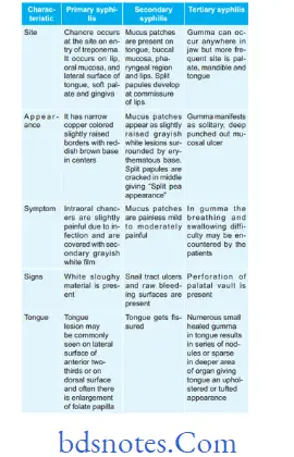
Question.1. Write notes on oral manifestations of syphilis.
Answer. Oral manifestations of syphilis are given below:
Question.2. Describe oral manifestations of leprosy, tuberculosis and syphilis.
Answer.
Oral Manifestations Of Leprosy
- In oral cavity, the disease produces tumor-like lesions called “lepromas” which are found on lips, gingiva, tongue and hard palate.
- Oral lesion appears as yellowish soft or hard sessile growth which have tendency to break down and ulcerate.
- Ulceration, necrosis and perforation of palate.
- Fixation of palate with loss of uvula.
- Difficulty in swallowing and regurgitation.
- Cobblestone appearance of tongue with loss of taste sensation.
- Chronic gingivitis, periodontitis and candidiasis are present.
- Enamel hypoplasia of teeth, pinkish discoloration of teeth and tapering of teeth is present.
Read And Learn More: Oral Pathology Question And Answers
Oral Manifestations Of Tuberculosis
- Tuberculous infection in oral cavity may produce nodules,vesicles, fisures, plaque, granulomas or verrucal papillary lesions.
- Tuberculous lesions of oral cavity are tuberculous ulcers,tuberculous gingivitis and tuberculosis of salivary gland.
- Tongue is most common location for the occurrence, besides this palate, gingiva, lips, buccal mucosa, alveolar ridge and vestibules may also be affcted.
- Tongue lesion: Tuberculous lesion of tongue develops on the lateral borders and appears as single or multiple ulcers which are well defied, painful, fim and yellowish gray in color.
- Lip lesions: Lesions produce small, nontender, granulating ulcer at mucocutaneous junction.
- Gingival lesions: These lesions produce small granulating ulcers with concomitant gingival hyperplasia.
- Tuberculous lesion of jaw bone: Chronic osteomyelitis of maxilla and mandible may occur and infection reaches to bone via blood or root canal or extraction socket.
Tuberculous osteomyelitis of jaw bone produces pain,swelling, sinus or fitula formation.
Question.3. Write note on congenital syphilis.
Or
Write short note on congenital syphilis.
Answer. Congenital syphilis is a rare entity that occurs in children born of an infected mother.
- This condition occurs due to transplacental infection with T. palladium during fetal development.
- Deciduous teeth are less frequently affcted as compared to the permanent teeth.
Features of Congenital syphilis
- Mulberry molars and screw driven shaped incisors occur due to involvement of developing tooth germs.
- Rhagades, i.e. fisuring and scaring at the corners of the mouth.
- Saddle nose or bull dog appearance is seen.
- There is mandible prognathism and increased inter dental spaces.
- Delayed eruption of teeth is present.
- Hypodontia and enamel hypoplasia is seen.
- There is occurrence of Hutchinson’s triad which consists of:
- Hypoplasia of incisors and molar teeth
- 8th nerve deafness
- Interstitial keratitis of eyes.
Treatment
High doses of penicillin are given.
Question.4. Write note on NOMA.
Answer. It is also called as gangrenous stomatitis.
It is rapidly spreading gangrene of oral and facial tissues occurring usually in debilitated or nutritionally deficient patients.
Predisposing Factors
- Occurs in undernourished persons.
- Debilitated from infections such as diphtheria, measles,pneumonia, scarlet fever,
- TB and blood dyscrasias.
- Excessive mechanical injury.
- It is a specific infection by Vincent’s organism.
Clinical Features
- It is seen chiefly in malnourished children.
- Common sites are areas of stagnation around the fied bridge or crown.
- Commencement of gangrene is denoted by blackening of skin. Small ulcers of gingival mucosa spread rapidly and involves surrounding tissue of jaw, lips and cheeks by gangrenous necrosis.
- Odor is foul and patient have high temperature.
- Overlying skin is inflamed, edematous and finally necrotic.
Treatment
Reconstructive surgery should be done along with palliative treatment.
Question.5. Describe histopathology of actinomycosis.
Or
Describe histologic features with diagram of actinomycosis
Answer. Actinomycosis is a chronic granulomatous suppurative and firosing infection. It is caused by filamentous,
Gram positive and anaerobic actinomycosis group of infections, i.e. Actinomycoses Israeli, Actinomycosis viscous, etc.
Histopathology
- Actinomycosis under microscope shows numerous abscesses whose centres are occupied by bacterial colonies.
- Bone tissue often exhibits extensive necrosis with multiple areas of granuloma formation.
- Bacterial colony consists of dense, eosinophilic masses of
Gram-negative filaments.
- Periphery of each colony shows clubshaped swellings and produces a “Ray fungus” like appearance.
- Colonies are surrounded by polymorphonuclear neutrophils followed by lymphocytes, plasma cells and multinucleated giant cells.
- Colonies are surrounded by the firous tissue wall at outer margin.
Question.6. Write note on tuberculosis.
Answer. Tuberculosis is a infectious granulomatous disease caused by Mycobacterium tuberculosis.
Pathogenesis
Clinical Features
- Patient suffers from episodic fever and chills, easy fatigability and malaise.
- There is gradual loss of weight with persistent cough with or without hemoptysis.
- Choroid tubercles are seen in children.
- Lupus vulgaris may occur in children.
Oral Manifestation
- Tongue is the most common site involved followed by palate, lips, buccal mucosa, gingiva and frenula.
- There is presence of irregular, superfiial or deep ulcer which is painful and will increase in size.
- There is presence of diffse hyperemic, nodular or papillary proliferation of gingival tissues.
- Tuberculous osteomyelitis occurs in the later stages of disease.
Histological Features
There is formation of granuloma exhibiting, foci of caseous necrosis surrounded by epithelioid cells, lymphocyte and occasionally multinucleated giant cells.
- Epithelioid cells are morphologically altered macrophages and appear like epithelial cells.
- Multinucleated giant cells are of LanghAns type.
- Area of caseous necrosis appears eosinophilic.
- Granuloma is surrounded by firous tissue and lymphocytes. At times dystrophic
- calcifiation is seen.
Treatment
Multidrug therapy (MDT) is recommended.
Question.7. Write oral manifestations of tuberculosis.
Or
Write short note on oral manifestations of tuberculosis.
Answer. Following are the oral manifestations of tuberculosis:
Tuberculous infection in oral cavity may produce nodules, vesicles, fissures, plaque, granulomas or verrucal papillary lesions.
- Tuberculous lesions of oral cavity are tuberculous ulcers, tuberculous gingivitis and tuberculosis of salivary gland.
- Tongue is most common location for the occurrence,besides this palate, gingiva, lips, buccal mucosa,alveolar ridge and vestibules may also be affcted.
- Tongue lesion: Tuberculous lesion of tongue develops on the lateral borders and appears as single or multiple ulcers which are well defied, painful and fim in consistency. Ulcers are deep and are painful.
Margins of ulcer are undermined with no induration.
Area surrounding the ulcer remains inflmed and edematous. Base of the ulcer is yellowish. - Lip lesions: Lesions produce small, nontender, granulating ulcer at mucocutaneous junction.
Gingival lesions: These lesions produce small granulating ulcers. Gingiva appear diffuse, hyperemic and nodular papillary proliferation is seen. - Tuberculous lesion of jaw bone: Chronic Osteomyelitis of maxilla and mandible may occur and infection reaches to bone via blood or root canal or extraction socket. Tuberculous osteomyelitis of jaw bone produces pain, swelling, sinus or fitula formation. As jaw bone is involved patient complains of swelling and diffilty in eating.
- Miliary lesion of oral mucosa in military tuberculosis is a small grey colored tubercle which breakdown and ulcerate.
- Periapical tissue can also be involved by the tubercle.
Tooth socket is filed with tuberculous granulation tissue and has small pink and red elevations.
Question.8. Write short note on actinomycosis.
Or
Write short essay on actinomycosis.
Answer. It is a chronic granulomatous suppurative disease which is caused by anaerobic or microaerophilic gram-positive non-acid fast-branched filamentous bacteria.
- The most common organism is Actinomyces israelii,A. naeslundii, A. viscosus, A. odontolyticus and A.propionica.
Classification of Actinomycosis Based on Location
- Cervicofacial: When there is involvement of face and cervical area
- Abdominal: When there is involvement of abdomen
- Pulmonary: When there is involvement of pleural cavity.
- Cutaneous: When there is involvement of skin
- Central: When there is involvement of bone
- Periphery: When there is involvement of soft tissue.
Pathogenesis
Disease originates when there is disruption of mucosal barrier which leads to invasion of bacteria. There is occurrence of initial acute inflmmation which is followed by chronic indolent phase.
Lesions appear as single or multiple indurations.
Clinical Features
Cervicofacial Actinomycosis
- Its occurrence is more common in males.
- Disease may remain localized to soft tissues or spread to involve salivary glands, bone or skin of face and neck.
- Most commonly involve area is submandibular region.
- Presence of trismus is there before formation of pus.
- The disease is characterized by presence of palpable mass which is indurated and is painless. Skin surrounding the lesion has wooden indurated area of firosis.
Abdominal Actinomycosis
- It is more severe form of disease.
- Patient complains of fever with chills and vomiting.
- There is involvement of liver and spleen.
- On palpation abdominal mass is felt which is the sign in diagnosis of disease.
Pulmonary actinomycosis
- Patient complains of fever with chills, cough and presence of pain in pleural cavity.
- Empyema is present and there is formation of sinus.
Treatment
Patient should be kept on high antibiotic therapy such as penicillin, cephalosporin, clindamycin, etc.
Question.9. Write short essay on pyogenic granuloma.
Or
Write short note on pyogenic granuloma.
Answer. Pyogenic granuloma is considered as an exaggerated conditioned response to minor trauma.
Pyogenic granuloma is a misnomer since condition is not associated with pus formation.
Etiology
- It is caused by microorganisms such as streptococci and staphylococci.
- If there is minor trauma to the tissue it provides the pathway for the non-specifi microorganisms which can cause pyogenic granuloma.
- Hormonal imbalance can lead to pyogenic granuloma.
- The sulfhdryl molecule is the agent which lead to pyogenic granuloma.
Clinical Features
- It occurs at the age of 10 to 40 years.
- Female predilection is present.
- Most affcted sits are lip, gingiva, tongue, palate, vestibule.
- Lesion is more common in maxillary anterior region.
- Lesion is elevated, pedunculated or sessile mass with a smooth, lobulated or warty surface which is ulcerated.
- On manipulation, the ulcer bleeds.
- Lesion is pink to red to purple in color depending on age of the lesion. It is usually painless and is soft in consistency.
- Size of the lesion ranges from 1 mm to centimeters.
Histopathology
- Overlying epithelium is thin and atrophic. At times, it is hyperplastic too.
- Surface of the epithelium is usually ulcerated and is replaced by thick firinopurulent membrane.
- Underlying connective tissue has number of endothelial lined vascular spaces engorged with RBCs and extreme proliferation of firoblasts and budding endothelial cells.
- There is presence of moderate infitration of PMN leukocytes, lymphocytes and plasma cells.
- Areas of hemorrhage and hemosiderin pigmentation is seen in connective tissue stroma.
Treatment
Surgical excision of the lesion is done.
Question.10. Write short note on pathogenesis of tuberculosis.
Answer. Following is the pathogenesis of tuberculosis:
Pathogenesis Of Tuberculosis
- The interaction of the bacilli and the host begins when droplet nuclei from infectious patients are inhaled.
- The majority of the bacilli are trapped and exhaled by ciliary action and a fraction less than 10% enters alveoli.
- In the initial stage of the hostbacterial interaction, either host’s macrophages control the multiplication of the bacteria or the bacteria grow and kill the macrophages.
- Non-activated monocytes attacted from bloodstream to the site by various chemotactic factors ingest the bacilli released from the lysed macrophages.
- Initial stages are asymptomatic; about 2-4 weeks after infection tissue damaging and macrophage activating responses develop.
- With the development of specifi immunity and accumulation of a large number of activated macrophages at the site of primary lesion, granulomatous reaction or tubercles are formed.
- The hard tubercle consists of epithelioid cells, LanghAns giant cells, plasma cells, and firoblasts. These lesions develop when host resistance is high.
- Due to cellmediated immunity in the majority of individuals, local macrophages are activated and lymphokines are released, which neutralize the bacilli and prevent further tissue destruction.
- The central part of the lesion contains caseous, soft, and cheesy necrotic material (caseous necrosis). This necrotic material may undergo calcification at a later stage called Ranne complex, in the lung parenchyma and hilar lymph nodes in few cases.
- Caseous necrotic material under goes liquefaction and discharges into the lungs leading to the formation of a cavity. Spontaneous healing of the cavity occurs either by firosis or collapse.
- Calcification of the cavities may occur in which bacteria persist.
- In early stages, the spread of infection is mainly by macrophages to lymph nodes, other tissues, and organs.
However, in children with poor immunity hematogenous spread results in fatal miliary TB or tuberculous meningitis.
Question.11. Enumerate bacterial lesion involving oral cavity. Describe clinical features, investigations, histopathology and management of tuberculosis.
Answer.
Enumeration Of Bacterial Lesions Involving Oral Cavity
- Syphilis
- Nonvenereal treponematoses
- Gonorrhea
- Streptococcal tonsillitis and pharyngitis
- Leprosy
- Tuberculosis
- Actinomycosis
- Noma
- Scarlet fever
- Diphtheria
- Cat scratch disease
- Tularemia
- Tetanus
- Rhinoscleroma
- Botryomycosis
- Melioidosis
- Granuloma Inguinale
- Lymphogranuloma venereum
- Myiasis.
Investigations
- Serology: In this ELISA technique is used which helpful in diagnosis of tuberculosis in children. PCR technique is more specifi and sensitive serological test than ELISA,but PCR is less used due to its high cost.
- Chest X-ray: Presence of multiple nodular infiltrations or ill-defied opacities in one of upper lobes is characteristic for pulmonary tuberculosis.
An area of translucency in radiopacities is indicative of cavitation. Presence of cavity is indicative of an active lesion.
In some of the patients, multiple thickwalled cavities can be seen. At the time of firosis, trachea and mediastinum shift to same side.
Fibrosis can also cause calcification. - Pathological tests:
- Blood examination: Peripheral blood examination shows monocytosis, i.e. 8 to 12%
- ESR is elevated.
- Tuberculin test: It is a test to recognize prior tubercular infection, and is done by injecting one unit of purifid protein derivative (PPD) on the forearm and readings taken after 48 hours. Induration of more than l5 mm indicates a positive test.
The younger is the patient, greater is signifiance of positive test.
A negative test does not always exclude tubercular infection since it may be negative in patients of blood malignancies, malnourishment and those on immunosuppressive therapy.
Tuberculin test is nonspecific and only indicates prior infection. Its sensitivity wanes with age.
Management
Chemotherapy.
Drugs for primary Chemotherapy
(First Line Anti-Tubercular Drugs)
Under Dots following treatment regimen is used
Second-line anti-tuberculous Drugs
Treatment regimen under rNTCP for MDr-TB (Multi- drug-resistant TB) and XDr-TB (Extensively drug-resistant TB)
- For MDR-TB:
- Six drugs in intensive phase for 6–9 months: Kanamycin,levoflxacin, ethionamide, cycloserine, pyrazinamide and ethambutol.
- Four drugs in continuation phase for 18 months:
Levofloxacin, ethionamide, cycloserine and ethambutol. - Reserve drug is paminosalicylic acid.
- For XDR-TB:
- Seven drugs in intensive phase for 612 months:
Capreomycin, p-aminosalicylic acid, moxiflxacin,high dose isoniazid, clofazimine, linezolid, Amoxicillin and clavulanic acid. - Six drugs in continuation phase for 18 months:
paminosalicylic acid, moxifloxacin, high dose lsoniazid, clofazimine, linezolid, amoxicillin and clavulanic acid.
Reserve drugs: Clarithromycin, thiacetazone
- Seven drugs in intensive phase for 612 months:
- Corticosteroids: They are to be given in the severe cases to enable them to survive till antitubercular drugs become effctive. Oral prednisolone is given in doses of 20 mg orally for 6 to 8 weeks. Steroids produce euphoria and increase appetite in the patients.
- Surgery: Surgical resection of infected lobe is feasible.
- Symptomatic treatment:
- Cough: If it is irritative, linctus codeine is given.
Smoking should be stopped - Laryngitis: Rest is given to the voice. If pain is present
anesthetic powders, spray and lozenges are given.
- Cough: If it is irritative, linctus codeine is given.
Leave a Reply