• Skip to main content
  • Skip to secondary menu
  • Skip to primary sidebar
  • About Us
  • Terms of Use
  • Privacy Policy
  • Disclaimer
  • Contact Us
  • Sitemap

BDS Notes

BDS notes, Question and Answers

  • Public Health Dentistry
  • Periodontics
  • Pharmacology
  • Pathology
  • Orthodontics
    • Anchorage In Orthodontics
    • Mandibular Growth, Functional Matrix
    • Retention and Relapse
  • General Surgery
    • Cysts: Types, Causes, Symptoms
    • Maxillofacial Fractures, Disorders, and Treatments
    • Lymphatic Disorders
    • Neurological and Facial Disorders
  • Temporal And Infratemporal Regions
    • Spinal and Neuroanatomy
  • Dental Materials
    • Dental Amalgam
Home » Arterial Anastomosis Of The Elbow

Arterial Anastomosis Of The Elbow

November 1, 2025 by Joankessler parkland Leave a Comment

Arterial Anastomosis Of The Elbow

Question 1. Write a short note on the arterial anastomosis around the elbow joint.
Answer.

The arterial anastomosis around the elbow joint is formed between the branches of the following arteries:

  • Brachial artery
  • Radial artery
  • Ulnar artery

Upper Limb Arterial Anastomosis Elbow Joint

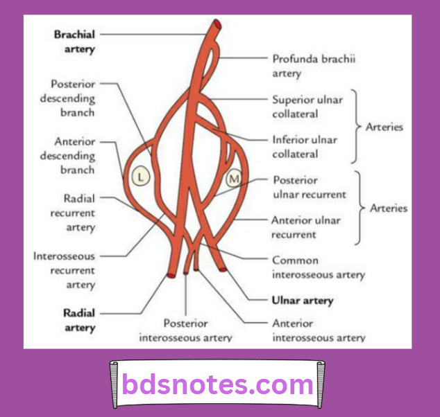
Arterial anastomosis of the elbow

Question 2. Write a short note on the cubital fossa.
Answer.

The cubital fossa is a triangular hollow in front of the elbow joint.

Cubital Fossa Anatomy Boundaries Contents

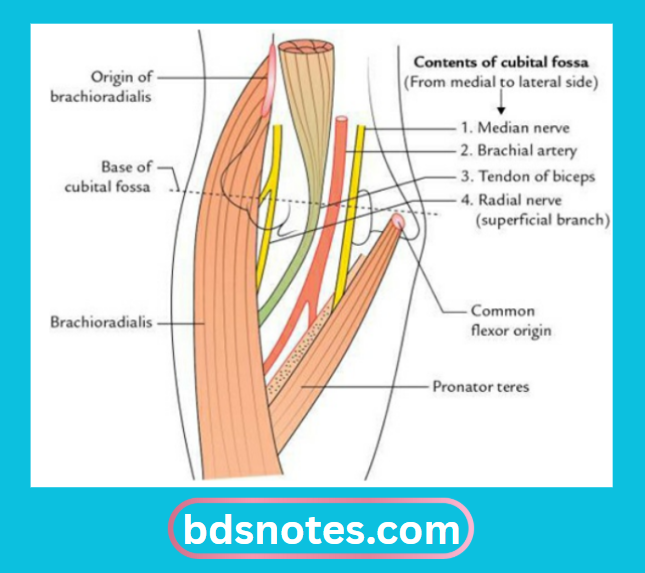
Cubital Fossa Boundaries

  • The base is formed by an imaginary horizontal line joining the medial and lateral epicondyles of the humerus.
  • The medial wall is formed by the pronator teres.
  • The lateral wall is formed by brachioradialis.
  • The roof is formed by skin, superficial fascia, deep fascia, and bicipital aponeurosis.
  • The superficial fascia contains the median cubital vein, the lateral cutaneous nerve of the forearm, and the medial cutaneous nerve of the forearm.
  • The floor is formed by the brachialis muscle in the upper and medial part, and the supinator muscle in the lower and lateral part.
  • Apex is a point where the pronator teres disappears underneath the brachioradialis muscle.

arterial anastomosis of elbow

Cubital Fossa Contents

From medial to lateral side:

  • Median nerve
  • Brachial artery
  • Biceps tendon
  • Superficial branch of the radial nerve
    Mnemonic: MBBS.

elbow joint anastomosis

Cubital Fossa Applied Anatomy

  • The brachial artery is auscultated in the cubital fossa for recording the blood pressure.
  • The median cubital vein is used in the region of the cubital fossa for venipuncture, as it lies superficial to the bicipital aponeurosis and is the most fixed vein.

Filed Under: Upper Limb

Reader Interactions

Leave a Reply Cancel reply

Your email address will not be published. Required fields are marked *

Primary Sidebar

Recent Posts

  • Branchial Cleft Cyst: Background, Pathophysiology, Etiology
  • Maxillary Nerve: Origin, Course, And Branches
  • The Father Of Anatomy And A Great Anatomist Herophilus
  • Bone Structure – Anatomy
  • The External Carotid Artery: Anatomy, Branches, And Functions
  • Occipitofrontalis Muscle
  • Superficial Temporal Artery
  • Platysma Muscle
  • Cartilage
  • Cauda Equina And Conus Medullaris Syndromes
  • Subcutaneous Injections And Device Management
  • Types Of Circulation: Pulmonary, Systemic, And Portal
  • Structure Of Skeletal Muscle
  • Elastic Cartilage
  • Cellular Organelles And Structure
  • The Golgi Apparatus – The Cell
  • The Cytoplasmic Inclusions Of Certain Plant Cells
  • Dental Abscess
  • Laser Surgery
  • Our Facial Muscles And Their Functions

Copyright © 2026 · Magazine Pro on Genesis Framework · WordPress · Log in