• Skip to main content
  • Skip to secondary menu
  • Skip to primary sidebar
  • About Us
  • Terms of Use
  • Privacy Policy
  • Disclaimer
  • Contact Us
  • Sitemap

BDS Notes

BDS notes, Question and Answers

  • Public Health Dentistry
  • Periodontics
  • Pharmacology
  • Pathology
  • Orthodontics
    • Anchorage In Orthodontics
    • Mandibular Growth, Functional Matrix
    • Retention and Relapse
  • General Surgery
    • Cysts: Types, Causes, Symptoms
    • Maxillofacial Fractures, Disorders, and Treatments
    • Lymphatic Disorders
    • Neurological and Facial Disorders
  • Temporal And Infratemporal Regions
    • Spinal and Neuroanatomy
  • Dental Materials
    • Dental Amalgam
Home » Applied Anatomy of the Nasal Septum: Little’s Area and Clinical Significance

Applied Anatomy of the Nasal Septum: Little’s Area and Clinical Significance

February 6, 2026 by Kristensmith Taylor Leave a Comment

Applied Anatomy of the Nasal Septum: Little’s Area and Clinical Significance

Describe gross anatomy of nasal septum. (or) Describe nasal septum. Give its blood supply, nerve supply, lymphatic drainage & applied anatomy (or) Nerve supply of septum of nose (or) Nasal septum
Answer:

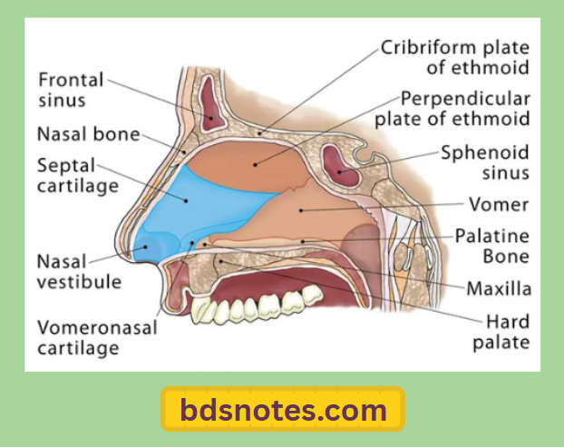
  • It is a median osseocartilagenous partition between the two halves of the nasal cavity

Nasal Septum Formation:

  • It is partly formed by bone & partly by cartilage
    1. Bony part
      • It is formed by
        1. Perpendicular plate of ethmoid
        2. Vomer
        3. Accessory bones like
          • Nasal spine of frontal bone
          • Sphenoidal crest & rostrum
          • Palatine processes of maxillae & horizontal parts of palatine bone
    2. Cartilages are formed by
      • Septal cartilage
      • Septal process of lower nasal cartilage
      • Vomeronasal cartilage

Nasal Septum  Blood supply, nerve supply & lymphatic drainage:

Paranasal Sinuses Vascular And Nerve Supply

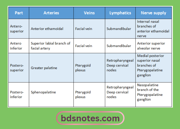
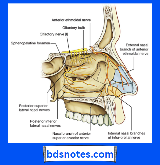
  • General sensory nerves arises from trigeminal nerve
  • Special sensory nerves are olfactory nerves which are confined to the upper part of olfactory area

Nasal Septum  Applied anatomy:
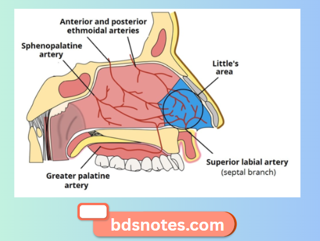
Little’s Area:

  • It is common site of epitaxis
  • It is an area of formation of large capillary network called the Kiesselbach’s plexus
  • It is formed by anastomosis of five arteries
    1. Sphenopalatine artery
    2. Superior labial branch of facial artery
    3. Anterior ethmoidal artery
    4. Greater palatine artery
    5. Nasal branches of facial artery
      • Pathological deviation of the nasal septum causes repeated attacks of common cold, allergic rhinitis, sinusitis, etc.

Nasal Septum Bones And Cartilage

Nasal Septum Arterial Supply Diagram

Nasal Septum Nerve Innervation Details

Filed Under: Anatomy

Reader Interactions

Leave a Reply Cancel reply

Your email address will not be published. Required fields are marked *

Primary Sidebar

Recent Posts

  • Branchial Cleft Cyst: Background, Pathophysiology, Etiology
  • Maxillary Nerve: Origin, Course, And Branches
  • The Father Of Anatomy And A Great Anatomist Herophilus
  • Bone Structure – Anatomy
  • The External Carotid Artery: Anatomy, Branches, And Functions
  • Occipitofrontalis Muscle
  • Superficial Temporal Artery
  • Platysma Muscle
  • Cartilage
  • Cauda Equina And Conus Medullaris Syndromes
  • Subcutaneous Injections And Device Management
  • Types Of Circulation: Pulmonary, Systemic, And Portal
  • Structure Of Skeletal Muscle
  • Elastic Cartilage
  • Cellular Organelles And Structure
  • The Golgi Apparatus – The Cell
  • The Cytoplasmic Inclusions Of Certain Plant Cells
  • Dental Abscess
  • Laser Surgery
  • Our Facial Muscles And Their Functions

Copyright © 2026 · Magazine Pro on Genesis Framework · WordPress · Log in