Angles Class 3 Malocclusion
Define Angle’s class 3 malocclusion. Differentiate between true and pseudo class 3 and write in detail the clinical features of class 3.
Or
Write difference between true and pseudo class 3.
Or
Differentiate between briefly true and pseudo class 3 malocclusion
Or
Write about clinical features of Angle’s Class 3 malocclusion.
Answer. It is defied as “a class 3 molar relationship refers to a condition where the mesiobuccal cusp of upper fist permanent molar occludes between the mandibular first and second molars”.
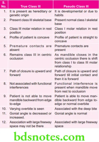
Difference Between True and Pseudo Class 3 Malocclusion
“Understanding the role of Angle’s Class 3 malocclusion in orthodontics”
Clinical Features/Clinical Picture of Class 3 Malocclusion
Following are the clinical features of class 3 malocclusion
Occlusal Features
- Molar relation is class 3, i.e. mesiobuccal cusp of upper first permanent molar occludes between the mandibular first and second molars.
- Canine relation is class 3, i.e. maxillary canine occludes in between mandibular fist and second premolars.
- Incisor relationship is class 3 with reverse overjet.
- Due to transverse relationship of arches posterior crossbite is seen.
- Maxillary arch is frequently narrow while the mandibular arch is broad. Posterior crossbite is a common feature.
- Maxillary teeth are crowded as arch is narrow and short in some cases.
- As chin is prominent, patient has concave profie.
- Vertical growers exhibit increased inter-maxillary height and may have an anterior open bite. In some patients deep overbite can be seen.
“Importance of studying Angle’s Class 3 malocclusion for better treatment outcomes”
Skeletal Features
- Maxilla is retrognathic.
- Mandible is prognathic.
- Combination of prognathic mandible and retrognathic maxilla is seen.
- Incisor, canine and molar relations are class 3
- Mentolabial sulcus is shallow.
- Chin is prominent.
- Lower facial height is increased.
“Common challenges in diagnosing and managing Angle’s Class 3 malocclusion”
Soft Tissue Features
- Facial profile is concave.
- Anterior facial divergence is present.
- Lips are incompetent.
- Short upper lip
- Tongue is anteriorly placed.
“Steps to identify common causes of Angle’s Class 3 malocclusion”
Functional Features
- Mandible is displaced forwardly.
- As there is unilateral crossbite, lateral mandibular displacement is present.
- In pseudo Class 3, patients have skeletal class 1 pattern, this abnormality is due to tilting of teeth and due to forward path of closure.
“Role of genetics in causing Angle’s Class 3 malocclusion”
Growth
- Unfavorable facial growth is seen in Class 3 cases
- Tendency to open bite increases with vertical facial growth.
- Excessive horizontal growth gets worse with reverse overjet.
Leave a Reply