Anatomy Of Tympanic Membrane
Write a short note on the tympanic membrane.
Answer.
Tympanic Membrane Introduction
- The tympanic membrane or eardrum is a semitransparent oval membrane, which separates the external acoustic meatus from the middle ear cavity.
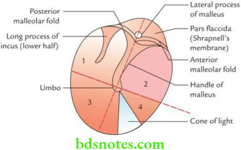
- Its diameter measures about 9 × 10 mm and is placed obliquely at an angle of 55° with the floor of the external acoustic meatus. It faces downwards, forwards, and laterally. The circumference of the membrane is made up of a fibrocartilaginous ring. The sulcus is absent between anterior and posterior malleolar folds. The part of the membrane-enclosed between malleolar folds is called pars flaccida.
“Importance Of The Tympanic Membrane In Hearing”
Tympanic Membrane Structure
Tympanic Membrane Structure consists of three layers:
- An outer cuticular layer (ectodermal in origin), continuous with the skin of the external auditory meatus.
- A middle fibrous layer (mesodermal in origin), consisting of superficial radiating fibers and deep circular fibers.
- An inner mucous layer (endodermal in origin), lined by ciliated columnar epithelium, continuous with the mucosa of the middle ear.
“Risk Factors For Damage To The Tympanic Membrane”
Tympanic Membrane Features
- Most of the tympanic membrane is tightly stretched and called pars tensa. A small upper part between two malleolar folds is loose and called pars flaccida (vide supra). The pars flaccida is crossed internally by the chorda tympani nerve.
- The tympanic membrane has outer and inner surfaces. The outer surface is concave. The inner surface is convex and provides attachment to the handle of the malleus, which extends up to its center. The point of maximum convexity on the inner surface is called the umbo. The cone of light is the reflection of light from the otoscope.
- The handle of the malleus is embedded in the middle fibrous layer.
“Early Signs Of Issues With The Tympanic Membrane”
“Understanding The Role Of The Tympanic Membrane In Hearing”
Tympanic Membrane Applied Anatomy
- The otoscopic examination may reveal the bulging, perforation, or retraction of the tympanic membrane.
- The membrane is incised (myringotomy) to drain the pus present in the middle ear.
- The incision should be given in the posteroinferior quadrant of the membrane to avoid injury to the chorda tympani nerve.
- The rupture of the tympanic membrane usually occurs in pars flaccida.
Leave a Reply