• Skip to main content
  • Skip to secondary menu
  • Skip to primary sidebar
  • About Us
  • Terms of Use
  • Privacy Policy
  • Disclaimer
  • Contact Us
  • Sitemap

BDS Notes

BDS notes, Question and Answers

  • Public Health Dentistry
  • Periodontics
  • Pharmacology
  • Pathology
  • Orthodontics
    • Anchorage In Orthodontics
    • Mandibular Growth, Functional Matrix
    • Retention and Relapse
  • General Surgery
    • Cysts: Types, Causes, Symptoms
    • Maxillofacial Fractures, Disorders, and Treatments
    • Lymphatic Disorders
    • Neurological and Facial Disorders
  • Temporal And Infratemporal Regions
    • Spinal and Neuroanatomy
  • Dental Materials
    • Dental Amalgam
Home » Anatomy Of Lumbrical Muscles In The Hand

Anatomy Of Lumbrical Muscles In The Hand

October 24, 2025 by Joankessler parkland Leave a Comment

Anatomy Of Lumbrical Muscles In The Hand

Question 1. Give the origin, insertion, nerve supply, and actions of lumbrical muscles.
Answer.

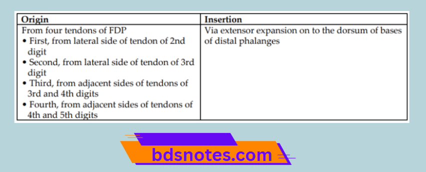
The origin and insertion of lumbrical muscles.

Lumbricals Flexor Tendon Relationship

“Understanding the anatomy of lumbrical muscles through FAQs: Composition, functions, and uses explained”

Origin and Insertion of Lumbrical Muscles

Lumbricals Flexor Digitorum Profundus Pathway

Lumbrical Muscles Nerve Supply

First and 2nd lumbricals are supplied by the median nerve, whereas 3rd and 4th lumbricals are supplied by the deep branch of the ulnar nerve.

“Importance of studying lumbrical muscles for medical students: Questions explained”

Lumbrical Muscles Actions

Flexion of metacarpophalangeal (MP) joints and extension of proximal and distal interphalangeal (PIP + DIP) joints.

Filed Under: Upper Limb

Reader Interactions

Leave a Reply Cancel reply

Your email address will not be published. Required fields are marked *

Primary Sidebar

Recent Posts

  • Branchial Cleft Cyst: Background, Pathophysiology, Etiology
  • Maxillary Nerve: Origin, Course, And Branches
  • The Father Of Anatomy And A Great Anatomist Herophilus
  • Bone Structure – Anatomy
  • The External Carotid Artery: Anatomy, Branches, And Functions
  • Occipitofrontalis Muscle
  • Superficial Temporal Artery
  • Platysma Muscle
  • Cartilage
  • Cauda Equina And Conus Medullaris Syndromes
  • Subcutaneous Injections And Device Management
  • Types Of Circulation: Pulmonary, Systemic, And Portal
  • Structure Of Skeletal Muscle
  • Elastic Cartilage
  • Cellular Organelles And Structure
  • The Golgi Apparatus – The Cell
  • The Cytoplasmic Inclusions Of Certain Plant Cells
  • Dental Abscess
  • Laser Surgery
  • Our Facial Muscles And Their Functions

Copyright © 2026 · Magazine Pro on Genesis Framework · WordPress · Log in