• Skip to main content
  • Skip to secondary menu
  • Skip to primary sidebar
  • About Us
  • Terms of Use
  • Privacy Policy
  • Disclaimer
  • Contact Us
  • Sitemap

BDS Notes

BDS notes, Question and Answers

  • Public Health Dentistry
  • Periodontics
  • Pharmacology
  • Pathology
  • Orthodontics
    • Anchorage In Orthodontics
    • Mandibular Growth, Functional Matrix
    • Retention and Relapse
  • General Surgery
    • Cysts: Types, Causes, Symptoms
    • Maxillofacial Fractures, Disorders, and Treatments
    • Lymphatic Disorders
    • Neurological and Facial Disorders
  • Temporal And Infratemporal Regions
    • Spinal and Neuroanatomy
  • Dental Materials
    • Dental Amalgam
Home » Anatomy And Histology Of The Pancreas

Anatomy And Histology Of The Pancreas

July 25, 2025 by Kristensmith Taylor Leave a Comment

Anatomy And Histology Of The Pancreas

Question 1. Histology of pancreas (or) Islets of Langerhans
Answer:

  • It is partly exocrine & partly endocrine gland
    1. Exocrine part
      • A delicate capsule surrounds the pancreas
      • Septa extends from capsule into the gland & divides it into lobules
        Secretory elements are long & tubular
      • The lining cells are triangular with spherical nuclei
      • Cytoplasm is basophilic & contains zymogen granules
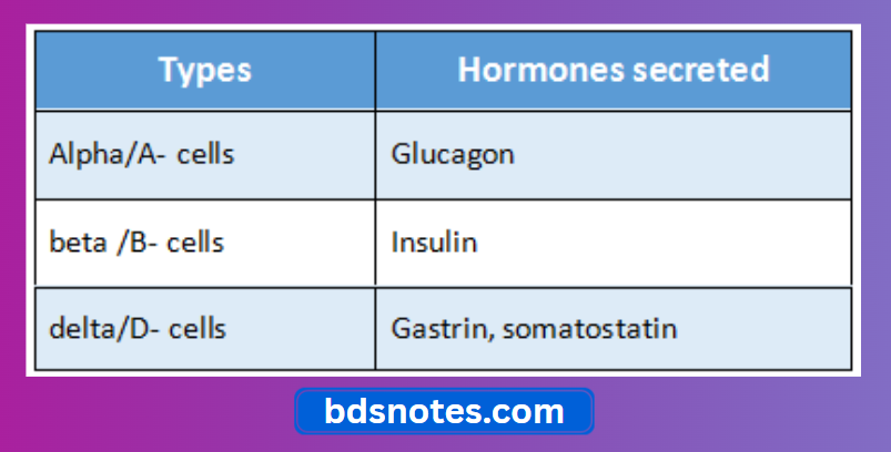
    2. Endocrine part
      • It consists of numerous collection of cells called islets of Langerhans
      • Each islets is separated from the surrounding alveoli by a thin layer of reticular tissue

“Understanding the pancreas through FAQs: Anatomy, histology, and functions explained”

Histology of pancreas Types:

Pancreatic Islet Cells And Hormones

“Importance of studying the anatomy and histology of the pancreas for medical students: Questions explained”

Question 2. Histology of Kidney
Answer:

Histology of kidney External structure:

  • It is a bean shaped structure
  • Its convex margin is placed laterally while concave margin is placed medially forming hilum
  • Hilum leads into renal sinus
  • Renal sinus is occupied by the upper expanded part of the ureter called renal pelvis
  • Renal pelvis divides into
    1. Major calyces
    2. Minor calyces
    3. Papilla

“Common challenges in mastering pancreas anatomy and histology notes effectively: FAQs provided”

Histology of kidney Internal structure:

  1. Kidney consists of
  2. Inner part called medulla 2 Outer part called cortex

Filed Under: Anatomy

Reader Interactions

Leave a Reply Cancel reply

Your email address will not be published. Required fields are marked *

Primary Sidebar

Recent Posts

  • Branchial Cleft Cyst: Background, Pathophysiology, Etiology
  • Maxillary Nerve: Origin, Course, And Branches
  • The Father Of Anatomy And A Great Anatomist Herophilus
  • Bone Structure – Anatomy
  • The External Carotid Artery: Anatomy, Branches, And Functions
  • Occipitofrontalis Muscle
  • Superficial Temporal Artery
  • Platysma Muscle
  • Cartilage
  • Cauda Equina And Conus Medullaris Syndromes
  • Subcutaneous Injections And Device Management
  • Types Of Circulation: Pulmonary, Systemic, And Portal
  • Structure Of Skeletal Muscle
  • Elastic Cartilage
  • Cellular Organelles And Structure
  • The Golgi Apparatus – The Cell
  • The Cytoplasmic Inclusions Of Certain Plant Cells
  • Dental Abscess
  • Laser Surgery
  • Our Facial Muscles And Their Functions

Copyright © 2026 · Magazine Pro on Genesis Framework · WordPress · Log in