Anatomy And Function Of The Temporomandibular Joint
Temporomandibular joint
Describe the temporomandibular joint (TMJ) under the following headings: (a) classification, (b) articular surfaces, (c) ligaments, (d) relations, (e) nerve supply, (f) movements and (g) applied anatomy.
Answer.
Temporomandibular Joint Classification
Temporomandibular Joint is synovial joint of condylar variety.
Special features of TMJ:
- Temporomandibular Joint is atypical synovial joint because its articular surfaces are covered by fibrocartilage instead of hyaline cartilage.
- Temporomandibular Joint is a complex synovial joint because its cavity contains an articular disc.
- Two condyles of mandible articulate with a mechanically single bony component, the cranium; hence, two TMJs together function as single unit and form a single craniomandibular joint of bicondylar variety.
“Understanding the anatomy and function of the TMJ through FAQs: Q&A explained”
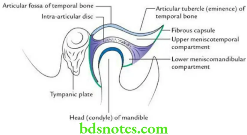
Temporomandibular Joint Articular surfaces
Above: Articular fossa and articular tubercle/eminence of temporal bone. It is concavoconvex from behind to forward and is covered by a fibrocartilage.
Below: Head of mandible. It is elliptical and is also covered by a fibrocartilage.
“Importance of studying the TMJ for dental and medical students: Questions explained”
The joint cavity is divided into two parts by an articular disc: (a) upper meniscotemporal compartment that permits gliding movements and (b) lower meniscomandibular compartment that permits rotational as well as gliding movements.
Temporomandibular Joint Ligaments
Temporomandibular Joint Main ligaments
- Capsular ligament: It is attached above to the articular tubercle, the circumference of mandibular fossa and squamotympanic fissure, and below to the neck of mandible.
- Lateral ligament/temporomandibular ligament: It is a thick band of fibrous tissue that covers the lateral aspect of capsule and strengthens it. It extends from articular tubercle on root of zygoma above to the lateral aspect of the neck of mandible below. Its fibres run downwards and backwards.

“Common challenges in understanding TMJ anatomy effectively: FAQs provided”
Temporomandibular Joint Accessory ligaments
- Stylomandibular ligament: It extends from styloid process of temporal bone to the angle of mandible. It is formed due to thickening in the investing layer of deep cervical fascia.
- Sphenomandibular ligament: It extends from spine of sphenoid to the lingula of mandible. It is derived from the first pharyngeal arch cartilage.
Temporomandibular Joint Relations
Temporomandibular Joint Lateral
- Skin and fasciae
- Parotid gland
- Temporal branches of facial nerve
Temporomandibular Joint Medial
- Tympanic plate of temporal bone
- Spine of the sphenoid and sphenomandibular ligament
- Auriculotemporal and chorda tympani nerves
- Middle meningeal artery
“Factors influencing success with TMJ knowledge: Q&A”
Temporomandibular Joint Anterior
- Lateral pterygoid
- Masseteric nerve and artery
Temporomandibular Joint Posterior
- Parotid gland, which separates the joint from the external auditory meatus
- Superficial temporal vessels
- Auriculotemporal nerve
Temporomandibular Joint Superior
- Middle cranial fossa
- Middle meningeal vessels
“Steps to explain the anatomy of the TMJ: Articular disc vs condyle vs ligaments: Q&A guide”
Temporomandibular Joint Inferior
- Maxillary artery and vein
Temporomandibular Joint Nerve supply
- Auriculotemporal nerve
- Masseteric nerve
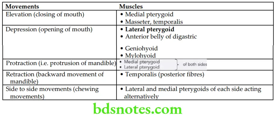
Temporomandibular Joint Movements
The movements of the temporomandibular joints and muscles producing.
Movements of TMJ and Muscles Producing Them
“Role of the articular disc in TMJ function: Questions answered”
Temporomandibular Joint Applied anatomy
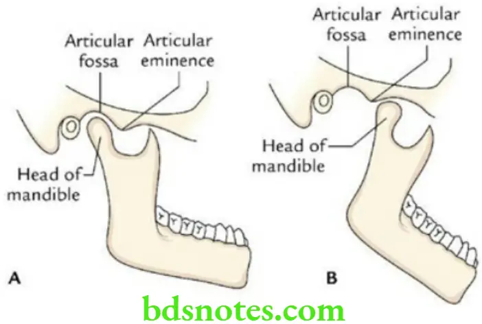
- Dislocation of temporomandibular joints (TMJs): The TMJs are mostly dislocated anteriorly. When the mouth is open, the mandibular condyles lie underneath the articular eminences of the temporal bone (the most unstable position of TMJ). In this position, if mouth is opened widely or even a severe muscular spasm (e.g. a convulsive yawn) it may displace the heads of mandible forward and upwards to be locked into the infratemporal fossa, leading to anterior dislocation of TMJ. As a result, there is inability to open mouth.
The reduction of joint can be easily achieved by pressing the molar teeth downwards with thumbs, and at the same time pushing the chin upward and backward. - Jaw clicking: The articular disc of TMJ may become partially detached from the capsule. As a result, movements of jaw becomes noisy and produces an audible click during movements of the TMJ.
“How does the TMJ enable chewing and speech? FAQ explained”
“Early warning signs of gaps in understanding TMJ basics: Common questions”
Articular Disc of TMJ
Articular Disc of Temporomandibular Joint General features
- It is an oval plate of fibrocartilage, which divides the cavity TMJ into two compartments: (a) an upper meniscotemporal compartment and (b) a lower meniscomandibular compartment.
- It presents a thick peripheral margin and a thin central part.
- It has concavoconvex superior surface and a concave inferior surface.
- Its periphery is firmly attached to the fibrous capsule.
- Morphologically, it represents the tendon of lateral pterygoid muscle.
“Differential applications of hinge vs sliding movements in the TMJ: Questions answered”
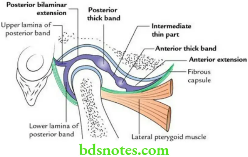
Parts of articular disc of Temporomandibular Joint
In sagittal section, it presents five parts. From before to backwards these are:
- Anterior extension
- Anterior thick band
- Intermediate thin part
- Posterior thick band
- Posterior bilaminar extension
“Asymptomatic vs symptomatic effects of ignoring TMJ principles: Q&A”
Leave a Reply