Causes of Agranulocytosis
“What is agranulocytosis? A detailed question and answers guide”
Agranulocytosis Causes
1. Endocrinal causes like hyperpituitarism, hypoadrenalism.
2. Agranulocytosis due to drugs: This is a very important cause and is related to dosage of drugs as well as sensitivity reaction. The main drugs are:
- Anticancer drugs
- Anti-inflmmatory drugs
- Phenothiazines and tranquilizers
- Sulfonamides and cotrimoxazole
- Antithyroid drugs
- Antidiabetic drugs
- Antihistaminics
- Antiepileptic drugs
- Antimicrobial agents
“Understanding agranulocytosis through FAQs: Causes, diagnosis, and management explained”

“Importance of studying agranulocytosis for healthcare professionals: Questions explained”
Read And Learn More: General Medicine Question And Answers
Agranulocytosis Diuretics:
- Defiiency of vitamin B12 and folate
- It is caused due to obliteration of bone marrow due to myelofirosis, lymphoma and sarcoma.
It is caused due to bone marrow damage due to Xray radiation. - Due to viral infections, i.e. hepatitis, HIV, inflenza, EBV
- Due to bacterial infections, i.e. enteric fever, tuberculosis and gramnegative bacterial septicemia
- Due to protozoal diseases, i.e. malaria and Kalaazar.
- Due to autoimmune diseases i.e. SLE, chronic autoimmune neutropenia
- Congenital i.e. in Kostmann’s syndrome
“Common challenges in diagnosing and managing agranulocytosis effectively: FAQs provided”
“Factors influencing success with agranulocytosis knowledge: Q&A”
Agranulocytosis Clinical Manifestations
- Females are more commonly affcted than males
- Early manifestations of agranulocytosis are may be in form of sore throat or pain.
- In large number of cases ulceromembranous lesions appear on throat, tonsils, gum, tongue and genitalia
- These are often covered with grayish black exudates and may become gangrenous.
- Lymph gland generally cervical groups and in some, there are generalized lymphadenopathy
- Liver and spleen may become enlarged.
- As disease progresses severe toxemia develops and patient may go into shock.
“Role of diagrams in understanding agranulocytosis mechanisms: Questions answered”
Agranulocytosis Management
- Firstly, the cause is removable which is identifible.
- Secondly, the infection is controlled and patient is put on isolation ward and barrier nursing is done.
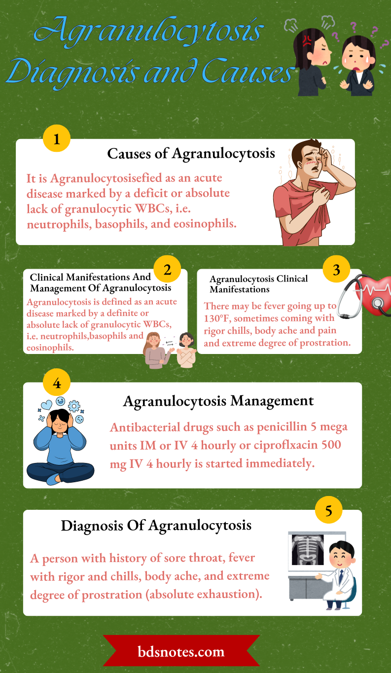
- Antibacterial drugs such as penicillin 5 mega units IM or IV 4 hourly or ciproflxacin 500 mg IV 4 hourly is started immediately.
- In addition metrogyl 500 mg every 6 hourly to take care of infection
- Anabolic steroids are also given.
- In cases where toxemia is severe corticosteroids are employed, i.e. injection dexamethasone 4 mg IV 6 hourly.
- Granulocytes transfusion is given to tide over crisis. This
is given daily for 5–7 days.
“Steps to master agranulocytosis for exams: Study plans vs mock tests: Q&A guide”
Diagnosis Of Agranulocytosis
The diagnosis of agranulocytosis is based on clinical history and physical examination.
- In large number of cases ulceromembranous lesions are present on throat, tonsil, gums, tongue and genitalia. The lesions are covered with grayish black exudates and become gangrenous.
- Confirmation of diagnosis is made by laboratory investigations.
- Peripheral blood fim shows complete absence of neutrophils.
- Bone marrow is hypocellular and there is depletion of myeloid elements.
Leave a Reply