Actinomycosis
Actinomycosis is a chronic granulomatous suppurative disease which is caused by anaerobic or microaerophilic gram-positive non-acid fast-branched filamentous bacteria.
- The most common organism is Actinomyces israelii,A. naeslundii, A. viscosus, A. odontolyticus and A.propionica.
Classification of Actinomycosis Based on Location
- Cervicofacial: When there is involvement of face and cervical area
- Abdominal: When there is involvement of abdomen
- Pulmonary: When there is involvement of pleural cavity.
- Cutaneous: When there is involvement of skin
- Central: When there is involvement of bone
- Periphery: When there is involvement of soft tissue.
“Importance Of Early Detection Of Actinomycosis”
Actinomycosis Pathogenesis
Disease originates when there is disruption of mucosal barrier which leads to invasion of bacteria. There is occurrence of initial acute inflmmation which is followed by chronic indolent phase.
Lesions appear as single or multiple indurations.
actinomycosis
Actinomycosis Clinical Features
Cervicofacial Actinomycosis
- Cervicofacial Actinomycosis occurrence is more common in males.
- Disease may remain localized to soft tissues or spread to involve salivary glands, bone or skin of face and neck.
- Most commonly involve area is submandibular region.
- Presence of trismus is there before formation of pus.
- The disease is characterized by presence of palpable mass which is indurated and is painless. Skin surrounding the lesion has wooden indurated area of firosis.
“Early Signs Of Actinomycosis Infection”
Abdominal Actinomycosis
- Abdominal Actinomycosis is more severe form of disease.
- Patient complains of fever with chills and vomiting.
- There is involvement of liver and spleen.
- On palpation abdominal mass is felt which is the sign in diagnosis of disease.
Pulmonary actinomycosis
- Patient complains of fever with chills, cough and presence of pain in pleural cavity.
- Empyema is present and there is formation of sinus.
cervicofacial actinomycosis
“Role Of Actinomyces Bacteria In Causing Actinomycosis”
Actinomycosis Treatment
Patient should be kept on high antibiotic therapy such as penicillin, cephalosporin, clindamycin, etc.
Actinomycosis
Actinomycosis is a chronic granulomatous suppurative and firosing infection. It is caused by filamentous,
Gram positive and anaerobic actinomycosis group of infections, i.e. Actinomycoses Israeli, Actinomycosis viscous, etc.
“Understanding The Role Of Actinomyces Bacteria In Actinomycosis”
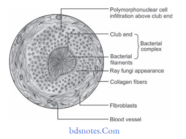
Actinomycosis Histopathology
- Actinomycosis under microscope shows numerous abscesses whose centres are occupied by bacterial colonies.
- Bone tissue often exhibits extensive necrosis with multiple areas of granuloma formation.
- Bacterial colony consists of dense, eosinophilic masses of
Gram-negative filaments.
“The Role Of Imaging In Diagnosing Actinomycosis Infections”
- Periphery of each colony shows clubshaped swellings and produces a “Ray fungus” like appearance.
- Colonies are surrounded by polymorphonuclear neutrophils followed by lymphocytes, plasma cells and multinucleated giant cells.
- Colonies are surrounded by the firous tissue wall at outer margin.
Leave a Reply