Abducens Nerve
Describe the abducent nerve under the following headings: (a) functional components, (b) origin, course and distribution and (c) applied anatomy.
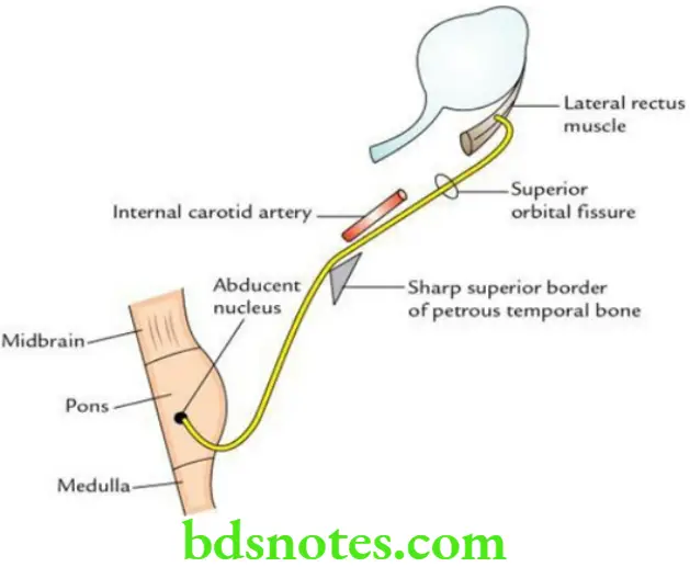
Answer. The abducent is CN 6. It is purely a motor nerve and supplies only one muscle – the lateral rectus of the eyeball.
“Factors influencing success with abducens nerve knowledge: Q&A”
Abducent Nerve Functional components
- General somatic efferent (GSE) fibres to supply lateral rectus. They arise from the abducent nucleus in the lower part of the pons.
- General somatic afferent (GSA) fibres carry proprioceptive sensation from the lateral rectus to the mesencephalic nucleus of the trigeminal nerve.
“Understanding the abducens nerve through FAQs: Q&A explained”
Abducent Nerve Origin, course and distribution The abducent nerve arises from the abducent nucleus in the lower part of the pons and emerges from the anterior surface of the brainstem at the junction of the pons and medulla oblongata. After emerging from the brain, it runs at first upwards, forwards and laterally in the posterior cranial fossa and pierces the dura mater over the clivus.
It then turns sharply forward, crossing the sharp superior border of the petrous temporal bone before entering the cavernous sinus. It traverses the cavernous sinus, lying at first lateral and then inferolateral to the internal carotid artery. The nerve enters the orbital cavity through the superior orbital fissure and supplies the lateral rectus muscle.
“Common challenges in understanding abducens nerve anatomy effectively: FAQs provided”
“Importance of studying the abducens nerve for medical students: Questions explained”
Abducent Nerve Applied anatomy The abducent nerve is a thin motor nerve that takes the longest intracranial course, and hence it is often damaged in increased intracranial pressure associated with coning of the brainstem.
The paralysis of the abducent nerve results in:
- Medial squint
- Diplopia
Leave a Reply