Protection From Radiation
Question 1. Write short note on protection from X-ray.
or
Describe briefly radiation protection of patient.
or
Discuss radiation protection of patient in detail.
Answer. The radiation protection of patient is describe as:
Read And Learn More: Oral Radiology Question And Answers
“Understanding the role of radiation protection in medical imaging: Q&A explained”
- X-ray machine: Good machines of reputed companies should be used.
- Selection of film: F- and E-speed films are used as they are of good quality and are highly sensitive. E speed films or Ekta speed films reduce exposure to 40%.
- Focal spot film distance: Longer is the focal spot film distance decrease is in the exposed tissue volume.
- Source skin distance: Increase in the source skin distance reduces the size of beam and reduces the volume of tissue irradiation which decreases the patient dose.
“Importance of studying radiation protection for better safety outcomes: Questions explained”
- Filtration: Low energy X-ray beam is removed by the filtration. As these X-rays do not contribute to the image formation they should be removed before they reach to the patient as they lead to the radiation exposure.
- X-ray collimation: It prevent the scattering. Beam should be collimated so that it is not more than 7 cm in diameter at the face of patient. Rectangular collimators should be preferred as they reduce the amount of tissue radiation.
- Intensifying screen: Use of rare earth screen decreases dosage for extraoral films.
- Grid: Grid decreases the fogginess of film due to the secondary radiation, this reduces the need for repeating the film.
- Kilovoltage: Operation of X-ray unit should be done at 60 to 90 kVp. X-ray beam of low kilovoltage leads to the higher patient doses, mainly to skin.
- Position-indicating devices: A 12 to 16 inches long position indicating device reduces exposure to patient as compared to short position indicating device. Open ended, circular or rectangular lead-lined cylinders are preferred to direct the X-ray beam.
- Lead aprons should be used who have lead content equivalent to 0.25 mm aluminum which is to be worn by patient during taking the radiograph.
Radiation Protection Exam Questions
- Thyroid collars should be weared to protect thyroid gland from radiation.
- Film-holding devices: They stabilize the X-ray film in mouth and so the hands of patient are not exposed to radiation.
- RVG: It decreases the dose of radiation required in IOPA.
“Common challenges in implementing radiation protection effectively: FAQs provided”
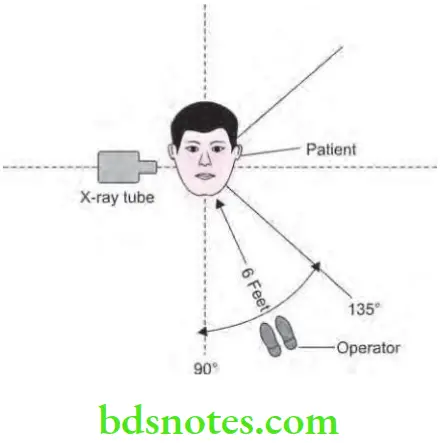
Question 2. Write short note on position and distance rule.
Answer.
- This rule implies on the protection of the operator.
- This rule is for the protection against primary beam.
- The rule states that the operator should stand at least 6 feet away from the source of radiation at an angle of 90 to 135° with respect to direction of central rays.
“Steps to explain the principles of radiation protection: ALARA vs shielding: Q&A guide”
- This rule takes advantage of inverse square law to reduce the intensity and also considers that in this position patient’s head will absorb most scattered radiation.
“Role of time, distance, and shielding in radiation protection: Questions answered”
Leave a Reply