Pathology Synopsis
General Pathology
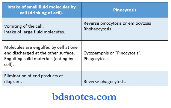
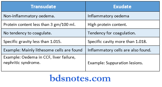
Some important definitions:
Abscess: A localized cavity containing purulent exudates or pus.
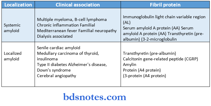
Amyloidosis is characterized by extracellular pathogenic fibrillar proteins that accumulate within the tissues and organs either because of increased synthesis or decreased catabolism.
Anaplasia: Lack of differentiation: It is a hallmark of malignant cells.
Angiogenesis: Occurs in inflammation, in which preexisting blood vessels give rise to capillary buds to produce new blood vessels
Aplasia: Absence of an organ because of failure of the developmental large to develop.
Apoptosis: Programmed cell death through activation of an internal suicide program to eliminate unwanted cells selectively with minimal disturbance to surrounding cells.
Pathology notes for medical students
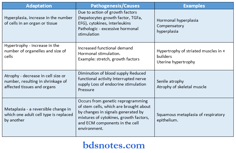
Atrophy: Shrinkage in the size of the cell and in the organ as a result of loss of cell shrinkage.
Cachexia: Extreme malnutrition with loss of body weight in cancer to AIDS condition.
Calcification: Abnormal deposition of calcium salts in soft tissues Spreading subcutaneous infection (mostly by strepto and staph).
An impaired cardiac function renders the heart unable to maintain an output sufficient for the metabolic requirement of the tissues and organs.
Bluish discoloration of mucus membrane due to the presence of reduced Hb. Repair of lost cells and tissues by connective tissue (fibroblasts).
This means inducing the arrest of bleeding at sites of vascular injury and maintaining blood in a fluid, clot-free state in normal vessels.
A state of metabolic equilibrium between the various organ of the body (by hormones). Overdevelopment of an organ due to an increased number of cells. Increase in organ size or function due to an increase in individual cell size. Corneal ulceration and eventual destruction of the cornea due to Vit A deficiency.
kernicterus: Increased un-conjugated bilirubin binds to lipids in the brain, causing – CNS damage.
leukoplakia: This is a pre-cancerous characterized by white patchy thickenings on the mucosa.
Metaplasia: A reversible change in which one adult cell type is replaced by another.
Metastasis: Invasion of the lymphatics, blood vessels, body cavities, etc by the tumor cells.
Moniliasis: (Candidiasis/thrush) – erythematous lesion on the palate and oral mucosa.
Neoplasia: A new growth due to a membrane of tissue formed as a result of abnormal, excessive, uncoordinated, autonomous, and purposeless proliferation of cells.
General pathology summary
Nephritic: Group of diseases resulting from increased permeability of the glomerular syndrome capillary wall and characterized by massive proteinuria, hyperalbuminemia, edema, hyperlipidemia, and hypercoagulability Cell injury – causes, responses, mechanism.
Pathology synopsis
Causes:
- O2 deprivation due to ischemia inadequate oxygenation, and loss of O2 – carrying capacity.
- Physical and chemical agents like trauma, cold, radiation, and drugs.
- Infectious agents
- Immunologic reactions
- Genetic derangements like chromosomal alteration
- Nutritional in balances
- Cellular response to injury depends on the type, duration, and severity of injury and adaptability of the cell, and accordingly, the cell adapts by undergo in atrophy, hyperplasia, hypertrophy, metaplasia, dysplasia, or maybe injured reversibly or irreversibly.
Vulnerable sites of injury are:
- Aerobic respiration involving ATP synthesis
- Cell membrane integrity
- Proteins synthesis (ribosomes)
- The integrity of genetic apparatus
Pathogenesis of cell injury:
- Decreased ATP synthesis, which leads to decreased protein synthesis, decreases.
- Na+ – K+ pump activity, decreased pH, and membrane injury.
- O2-derived free radicals cause membrane damage by lipid peroxidation, oxidation of protein, DNA damage, and cytoskeletal damage.
- Increased intracellular Ca2+, which activates phospholipases, proteases, ATP uses, and endonucleases.
- Direct membrane damage leads to defects in permeability
- Mitochondrial damage causes leakage of cytochrome into the cytosol and activation of the apoptotic pathways
- Free radicals – OH, H+, CCI3′, ONOO, NO2, NO3.
Neoplasia in pathology
Irreversible cell injury – Necrosis and Apoptosis:
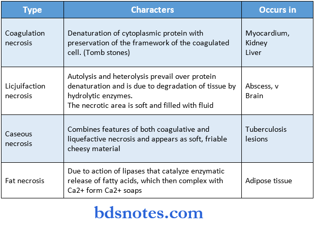
- Necrosis – cellular death due to denaturation of proteins and enzymatic digestion of organelles and other cytosolic components released by the cell.
Apoptosis- a form of programmed and coordinated cell death to eliminate unwanted cells selectively.
Example: Cell death during embryogenesis Cell death in tumors Cell death in cytotoxic T cells.
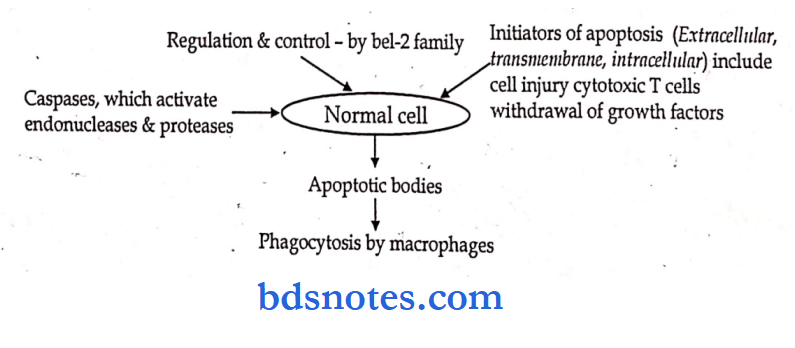
Apoptosis – a form of programmed and coordinated cell death to eliminate unwanted cells selectively.
Example: Cell death during embryogenesis Cell death in tumors Cell death in cytotoxic T cells
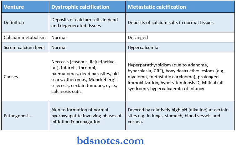
Calcification – abnormal deposition of Ca2+ salts in soil tissues.
Differences between dystrophic and metastatic calcification
Classification of amyloid:
Cellular adaptations:
Inflammation- reaction of vascularized tissue to local injury caused by nociceptive agents.
It is a body defense reaction in order to eliminate the injurious agent and can also, however cause life-threatening hypersensitivity reactions.
Classical signs – named by Celsus
- Rubor – (redness)
- Tumor (swelling)
- Color (heat)
- Dolor (pain)
- Function laksa (loss of function) by Virchow
MBBS pathology notes
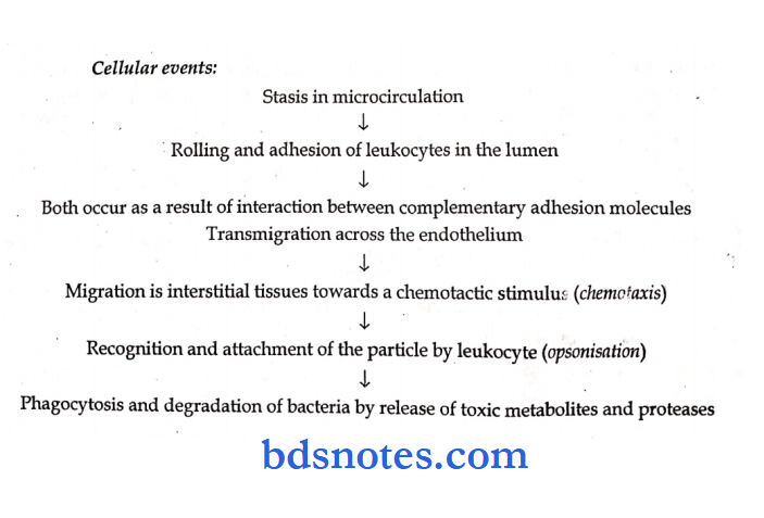
Events – pathogenesis
Vascular events:
- Initial transient vasoconstriction due to reflex spasm.
- Persistent progressive vasodilatation (due to vasodilator) causes increased blood flow (responsible for redness and warmth) and increased local hydrostatic pressure resulting in transudation of fluid into extracellular space (responsible for swelling).
- Stasis occurs due to increased vascular permeability arid leads to desmocytic margination.
Types of inflammation
Mechanism of increased vascular permeability:
- Endothelial cell contraction leads to widened intercellular gaps by histamine, and bradykinins.
- Endothelial cell retraction by cytokines, IL – 1d
- Direct endothelial injury resulting in necrosis and detachment by necrotizing agents
- Leukocyte-mediated injury due to the release of toxic oxygen species & proteolytic enzymes.
- Increased transcytosis.
- Leakage from regenerating capillaries.
Inflammatory cells

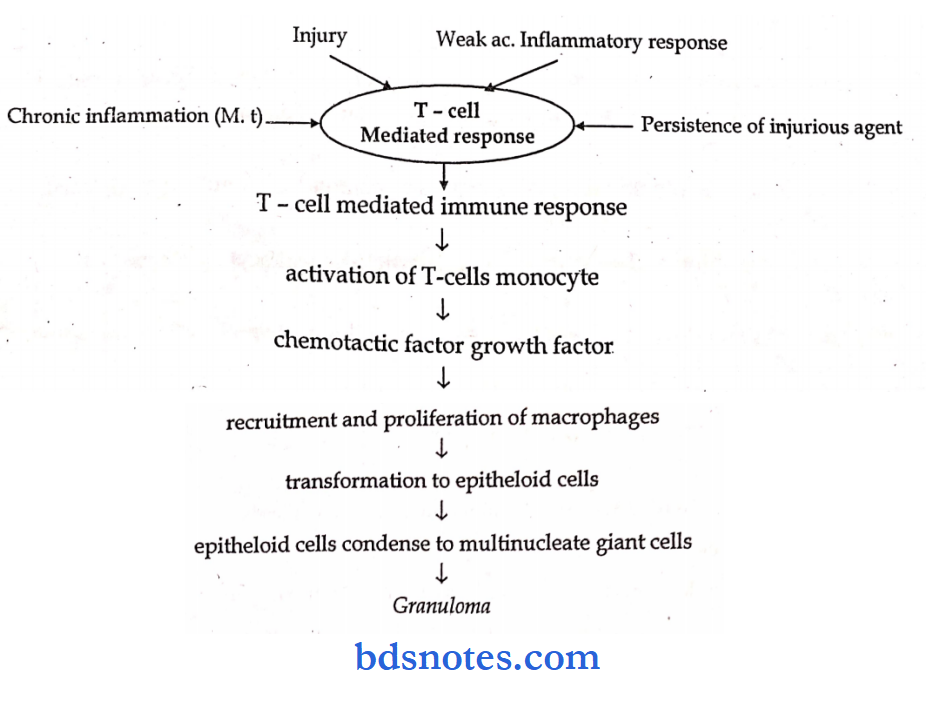
Granulomatous inflammation- is a distinctive chronic inflammatory reaction in which the predominant cell type is an activated macrophage with a modified epithelial-like appearance and is characterized by granuloma (focal collections of epitheloid macrophages that are surrounded by a collar of lymphocytes and occasionally plasma cells).
Pathology MCQs with answers
General Pathology
- The scavenger cells of the body are mononuclear phagocytes is macrophages.
- Generalized edema is called Anasarca or dropsy.
- The escapement of leucocytes from blood vessels in acute inflammation is called Emigration.
- The simultaneous escapement of R.B.C. along ‘with leucocytes in acute inflammation is called as Diapedesis.
- The emigration of leucocytes guided by chemical mediators is called Chemotaxis.
- Arachidonic add is formed horn essential fatty acid linoleic add.
- A decrease in the number of leucocytes in peripheral blood is called Leukopenia,
- An increase in the number of circulating leucocytes is called Leucocytosis
- Acute suppurative inflammation of hair follicles is called Boil or furuncle
- Loculated abscess in the tire dermis and soft tissue of the neck seen in untreated diabetics is Carbunde.
Systemic pathology overview
- A catarrhal inflammation is characterized by Mucous production.
- A suppurative inflammation is characterized by Pus production.
- The percentage of carcinomas arising at the lesser curvature of the stomach is more than 75%
- Analphlotoxins of the complement cascade are C3a and C5a.
- The half-life of monocyte is 1-2 days.
- The major stimulator of monocytes is Gamma interferon.
- Repair of the inflammatory process begins within hours 24 hours.
- The proliferation of epithelium around would margins occur within hours 48 hours.
- The major site for the maturation of B-lymphocytes is Bone marrow.
- A major site for maturation of T-lymphocyte is Thymus.
- A predominant lymphocyte in peripheral blood is T-lymphocyte.
- The predominant lymphocyte in the gingiva is B-lymphocyte.
- Vitamin, which maintains epithelial integrity, is Vit. A.
- Vitamins are necessary for proper development and function of bone Vit. A and Vit. D.
- Vitamin necessary for collagen formation and wound healing Vit. C.
- Macrophages are derived from cells’ Monocytes.
- Giant cells are derived from cells of Perivascular monocytes and macrophages.
- Localized inflammatory areas of fibrinoid necrosis are seen in the heart and are a pathognomic of rheumatic fever in Achoff bodies.
- Infarcts of the kidney are usually located in Pelvis-cortex.
- The most vulnerable organ to ischemia during shock is the Heart.
- Polyarteritis nodosa commonly affects the Kidney.
- The commonest site of metastatic calcifications is the Lungs.
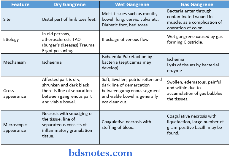
- Fat necrosis is common in Breast.
- The commonest site of liquefactive necrosis Brain.
- HLA – complex in humans is present on chromosome number 6
- Generalized edema of the body is called Anasarca,
- Edema of the peritoneal cavity is called Ascites.
- Squamous cell carcinoma developing from previous bum injuries is called Marjolin’s ulcer.
- The most favorable prognosis is shown by the type of Hodgkin’s disease Nodular sclerosis and lymphocytic predominant.
- The most common type of oral cancer is squamous cell carcinoma.
- The most common type of lung cancer is squamous cell carcinoma.
- The most common skin malignancy is Basal cell carcinoma.
- The hematogenous spread of malignancies is common in the organs Lung.
- A Tumour composed of multiple tissues representing all the 3 layers of an embryo is Teratoma.
- The disorganized proliferation of cells is common to their site of organ Hamartoma.
- A benign tumor arising from skeletal muscle is called Rhabdomyoma.
- A malignant tumor arising from skeletal muscle is called Rhabdomyo sarcoma.
- A benign neoplasm of blood vessels is called an Angioma.
- A malignant neoplasm of blood vessels is Angio sarcoma.
- A hard neoplasm is called Scirrhous.
- The definite diagnosis of neoplasm is by Histological Examination by biopsy.
- Hemorrhage through a permanent rupture in the vessel wall is called Rhexis.
- Variation in the size of R.B.C. is called Anisocytosis.
- Variation in the shape of R.B.C. is called Poikilocytosis.
- The strongest acid in the body is Heparin.
- “Cor bovine appearance of the heart is seen in Syphilis.
- Heberdens nodes are commonly seen in Osteoarthrosis.
- The commonest glycogen disorder is Vongierkes.
- Intracellular remnants of hemoglobin are called bodies Heinz.
- The commonest congenital coagulation disorder is Haemophilia – A.
- Leukemia associated with Philadelphia chromosome
Chronic myeloid leukemia. - Leukemia associated with young adolescents and children Acute lymphoblastic leukemia.
- ‘Starry-Sky’ appearance in the histological sections is seen in lymphoma Burkin’s lymphoma.
- ‘Reid-Sternberg’ cells are seen in Hodgkin’s lymphoma.
- Intra-nuclear dane particles or Australian antigen is found in the viral disease Hepatitis – B.
- Commonest leukemia is CLL.
- The commonest type of cell in CLL is B-Lymphocyte.
- Auer rods are present in type of leukemia acute myeloid leukemia.
- Gingival swellings are common in type of leukemia chronic lymphoblastic leukemia.
- The treatment of choice for aplastic anemia is Bone marrow transfer.
- The most common skin cancer is Basal cell carcinoma.
- The most common malignancy of bone is Osteosarcoma.
- The death in malignancy caused due to starvation is called Cachexia.
- The most common bone tumor in children is Ewing’s sarcoma.
- Commonest malignancy in children is Leukemia.
- The malignant neoplasm of bone which produces a sun-ray appearance on radiographs is Osteosarcoma.
- The malignant neoplasm which produces an onion-skin appearance as radiographs is Ewing’s sarcoma.
- The most common cancer death in females is Breast cancer.
- The type of nevus most commonly predisposes to malignant melanoma is Junctional nevus.
- In general malignant neoplasm arising from epithelium or glands is called Sarcoma.
- Carcinoma metastasis occurs by route Lymphatic.
- Sarcomas spread by the route Blood supply.
- Verocay bodies are present in neoplasm Neurolemmoma or Shwanomma.
- The abnormal proliferation of tissues that are native to that area is called a Hamartoma.
- Abnormal proliferation of tissues or structures that is not native to the area Christmas.
- Sarcoma which is spread by lymphatics is Rhabdomyosarcoma.
- Interstitial pneumonia is a common type of infection Viral.
- The most serious form of pneumoconiosis is Silicosis.
- The pneumoconiosis associated with bronchogenic carcinoma Asbestosis.
- The collection of pus in the lungs is called Empyema.
- Toxic injury of the brain in infants resulting from increased levels of unconjugated bilirubin is called Kernicterus.
- Jaundice becomes clinically evident when total serum bilirubin exceeds mg 100-ml/2 mg/dl
- Normal serum bilirubin concentration is 0.2 to 0.8 mg/100 ml
- The common cause of oesophageal varices seen in liver cirrhosis is Portal hypertension.
- Osteomyelitis is more commonly caused by the bacteria Staphylococcus aureus.
- In sickle cell anemia, osteomyelitis is commonly caused by the bacteria Salmonella.
- The presence of a significant number of non-multiplying bacteria in the blood is called Bacteremia.
- The presence of rapidly multiplying pathogenic bacteria in the blood is called Septicemia.
- Dissemination of small septic thrombi in the blood resulting in multiple abscesses or septic infarcts Pyemia.
- Granulomas in tuberculosis is called Tubercle.
- The fungal disease, which resembles tuberculosis in primary acute form Histoplasmosis.
- Tuberculosis of the lung is called Phthisis.
- Tuberculosis of cervical lymph nodes is Scrofula.
- Primary tuberculosis of the skin is called Lupus vulgaris.
- Infection mononucleosis is also called a disease since it spreads by saliva Kissing disease.
- A common cause of mycotic meningitis is Cryptococcus neoformans.
- The oral manifestation of advanced leishmaniasis is Cancrum oris.
- The most common infective organism in acute bacterial endocarditis is Staphylococci aureus.
- The most common infective organism in sub-acute bacterial endocarditis is Streptococcus viridans.
- Painful tender nodules on the fingertips of hands seen in SABE are called Osier’s nodes.
- Painless subcutaneous maculopapular lesions of the finger seen in acute bacterial endocarditis are called Jane Way’s pots.
- The hypertrophy of one paired organ following the defect of the other is called Vicarious hypertrophy.
- Potato nodes are seen in Sarcoidosis.
- Rubbery lymph nodes are characteristic of Lymphoma.
- Tuberculosis of the spine is called Pottis disease.
Definition and causes:
- Inflammation is defined as the local response of living mammalian tissues to injury due to any agent.
Signs of inflammation:
- Rubor (redness)
- Tumor (swelling)
- Calor (heart)
- Dolor (pain) and
- Functi laesa (loss of function).
The sequence of events in acute inflammation
- Vascular changes.
- Margination and pavements of Leucocytes.
- Emigration.
- Chemotaxis.
- Phagocytosis.
Systemic pathology synopsis
Stages of Phagocytosis
- Opsonization (or) attachment.
- Engulfment.
- Secretion or degranulation
- Degradation.
- Factors that cause edema formation Vascular factors
- Decreased plasma oncotic pressure (also called colloidal osmotic -pressure).
- Increased capillary Hydrostatic pressure.
- Increased capillary permeability.
- Tissue factors
- Increased oncotic pressure of the interstitial fluid.
- Decreased tissue tension.
- Lymphatic obstruction (or) decreased lymphatic drainage.
- Increased sodium and water retention.
- Factors, which prevent edema formation or retain fluids within vascular compartments.
- Increased blood colloidal osmotic pressure.
- Decreased capillary hydrostatic pressure.
- Decreased capillary permeability.
- Decreased tissue oncotic pressure.
- Increased tissue hydrostatic pressure (or) tissue tension.
- Increased lymphatic drainage.
Calcifications:
- Congestive cardiac failure
- Right-sided heart failure (corpulmonnk) results from pulmonary hypertension (or) pulmonary stenosis.
Features are:
- Systemic venous congestion.
- Congestion of spleen, liver kidney.
- Oedema of ankle and enlargement of liver and spleen.
- Dullness and confusion.
- Left-sided heart failure results from systemic hypertension or myocardial infarction (or aortic stenosis)
Features are:
- Chronic congestion of the lung.
- Broun indentation of the lung.
- Dyspnoea.
- Unconjugated hyperbilirubinemia
- Hemolysis.
- Failure to conjugate bilirubin as seen in trigger-najja’s syndrome and chloramphenicol
Conjugated hyperbilirubinemia
- Obstruction of bile stones due to impacted stones in the common bile duct or cancer of the head of the pancreas.
Failure of liver cells to secrete conjugated bilirubin is seen in cirrhosis and hepatitis.
- Hypersecretion of growth hormone in children Hypersecretion of growth hormone in adults Hypo thyroidism in children Hypo thyroidism in adults
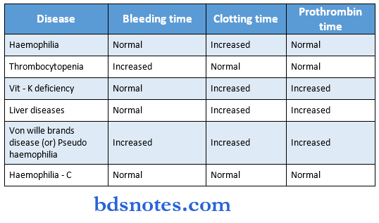
- Bleeding time and clotting time
Types Of Anaemia
- Microcytic hypochromic
- Iron deficiency anemia.
- Thalassemia.
- Sideroblastic anemia.
- Chronic blood loss.
- Nonnocytic normochromic
- Acute blood loss.
- Hemolytic anemia.
- Bone marrow failure.
- Macrocytic and hyperchromic
- Megaloblastic anemia due to Vitamin B12 folic acid deficiency.
Leave a Reply