Pindborg Tumor
“How does a Pindborg tumor form in the jaw?”
Answer. Pindborg tumor is locally aggressive neoplasm, which is also known as calcifying epithelial odontogenic tumor.
Pathogenesis Of Pindborg tumor
- Some investigators suggest that the Pindborg tumor arises from remnants of cells in stratum intermedium layer of the enamel organ in tooth development. Some hypothesize that the Pindborg’s tumor arises from the remnants of the primitive dental lamina.
- Definite etiology of neoplasm still remains enigmatic.
“Understanding Pindborg tumor: Causes and symptoms”
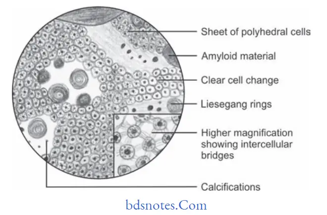
pindborg tumor
Clinical Features Of Pindborg Tumor
- Tumor occur in middle-aged persons.
- Mandible is involved more often than maxilla.
- Molar region is more common site of occurrence followed by premolar region.
- Tumor presents a slow enlarging, painless swelling of jaw with expansion and distortion of cortical plates.
- Swelling is bony hard and clinically, it is well defined or diffused.
- Pain, paresthesia may develop on rare occasions and few lesions may be completely asymptomatic.
“Importance of early diagnosis of Pindborg tumor”
calcifying epithelial odontogenic tumor
Histopathology Of Pindborg tumor
- Tumor reveals sheet of closely packed, polyhedral cells in noninflmed connective tissue stroma.
- Tumor cells contain ovalshaped nuclei and homogenous eosinophilic cytoplasm.
- Prominent intra cellular bridges and distinct cell boundaries are often found in the lesions.
- Some amount of homogenous, hyaline material is often deposited in between tumor cells called amyloid material.
- One of the most important histological characteristics of CEOT is the presence of several calcifid masses in and around the tumor cells.
- Some Liesegang rings are also found.
“Common features of Pindborg tumor explained”
“Calcifying epithelial odontogenic tumor (CEOT) explained”
Treatment Of Pindborg tumor
Surgical enucleation is done.
Odontogenic Tumors Mcqs With Answers
Leave a Reply