Histopathology of Calcifying Odontogenic Cyst (COC): A Microscopic View
Question. Write a short note on the histopathology of COC.
Answer. It is also known as a calcifying odontogenic cyst or Calcifying epithelial odontogenic cyst.
“Understanding the role of histopathology in diagnosing COC: Q&A explained”
Histopathology Of COC
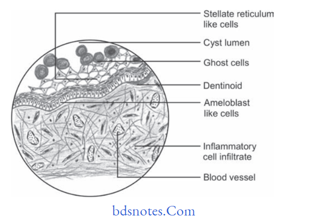
- Epithelial lining of COC shows a prominent basal cell layer consisting of palisaded columnar or cuboidal cells and hyperchromatic nuclei, which are polarized away from the basement membrane.
- Epithelium is 6–8 cell layers thick.
- Budding from the basal cell layer into adjacent connective tissue and epithelial proliferations into the lumen are seen.
“Importance of studying histopathology of calcifying odontogenic cyst: Questions explained”
“Common challenges in identifying COC effectively: FAQs provided”
- Ghost cells: They are enlarged, ballooned in shape, ovoid, or elongated elliptoid epithelial cells.
They are eosinophilic.
They are found in thick areas of the epithelial lining.
Cell outlines of these cells are well defined and at times they may be blurred. They are seen singly and also present in groups.
Few ghost cells also show nuclear remnants.
Ghost cells have an abnormal type of keratinization and have an affinity for calcification.
“Factors influencing success with COC diagnosis: Q&A”
- Ghost cells may also remain in contact with the connective tissue wall of the cyst, where they lead to foreign body reactions with the formation of multinucleated giant cells.
- An atubular dentinoid is also seen in the wall close to the epithelial lining and is also concerning epithelial proliferation.
- Dentinoids are found particularly in contact with masses of ghost cells.
Leave a Reply