Osteogenic Sarcoma: Clinical Features, Radiographic Stages, and Histopathology
Question. Defie neoplasm. Describe in brief clinical features, X-ray details and histopathology of osteogenic sarcoma.
Answer. “A neoplasm is an abnormal mass of tissue, the growth of which exceeds and is uncoordinated with that of the normal tissues and persists in the same excessive manner after cessation of the stimuli which evoked the change.” Willis
“Understanding osteogenic sarcoma through FAQs: Q&A explained”
Osteogenic Sarcoma is a common malignant neoplasm arising from the bone and beside plasma and myeloma.
Osteogenic Sarcoma Clinical Features
- It occurs during 10–25 years of age in the jaw and males are more commonly affected.
- The tumor involves maxilla more often than mandible.
- A very fast enlarging, painful swelling of jaw, causes expansion and distortion of cortical plates.
- Severe facial deformity and diffilty in taking the food due to restricted jaw movement.
- Displacement and loosening of regional teeth.
- Ulceration, hemorrhage and pathological fracture of bone are commonly associated.
“Importance of studying osteogenic sarcoma for better diagnostic outcomes: Questions explained”
Osteogenic Sarcoma X-ray Details
The Xray details of osteosarcoma are divided into following stages, i.e.
Osteogenic Sarcoma Osteolytic Stage
- It reveals moth eaten appearance.
- Border of the lesion at this stage are ill-defied.
- There is perforation and expansion of cortical plates.
- Lamina dura is absent, i.e. it get destroyed.
- Pathological fracture may be present.
- Root resorption is present.
Osteogenic Sarcoma Mixed Stage
- It is called as mixed because this stage show formation and destruction of bone.
- It reveals honeycomb appearance.
- Margins of lesion are ill defied.
“Common challenges in diagnosing osteogenic sarcoma effectively: FAQs provided”
Osteogenic Sarcoma Osteoblastic Stage
- It reveals sun ray appearance.
- At times subperiosteal bone laid down in layers which results in onion peel appearance.
- In osteosarcoma periosteum is elevated over the expanding tumor mass in a tent like fashion. At point on the bone where the periosteum begin to merge an acute angle between periosteum and bone is created which is known as
- Codman’s Triangle.
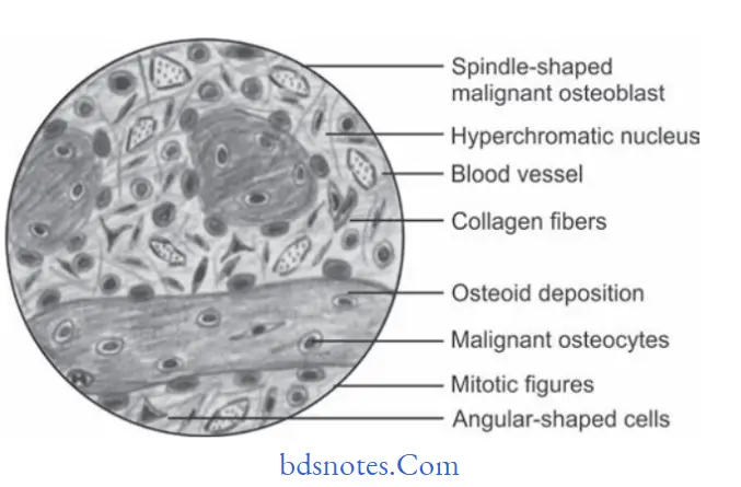
Osteogenic Sarcoma Histopathology
- There will be presence of numerous, actively proliferating,spindle shaped, oval or angular, malignant osteoblast cells within cellular stroma.
- The malignant osteoblast cells exhibit cellular pleomorphism, abnormal increased mitosis and nuclear hyperchromatism
- Multiple areas of newly formed bone or osteoid tissue are often present within firous stroma.
- In chondroblastic variations the malignant tumor cells produce large amount of cartilagenous tissue within tumor.
“Steps to explain causes of osteogenic sarcoma: Genetic mutations vs environmental factors: Q&A guide”
Leave a Reply