• Skip to main content
  • Skip to secondary menu
  • Skip to primary sidebar
  • About Us
  • Terms of Use
  • Privacy Policy
  • Disclaimer
  • Contact Us
  • Sitemap

BDS Notes

BDS notes, Question and Answers

  • Public Health Dentistry
  • Periodontics
  • Pharmacology
  • Pathology
  • Orthodontics
    • Anchorage In Orthodontics
    • Mandibular Growth, Functional Matrix
    • Retention and Relapse
  • General Surgery
    • Cysts: Types, Causes, Symptoms
    • Maxillofacial Fractures, Disorders, and Treatments
    • Lymphatic Disorders
    • Neurological and Facial Disorders
  • Temporal And Infratemporal Regions
    • Spinal and Neuroanatomy
  • Dental Materials
    • Dental Amalgam
Home » Special Stains in Histopathology: Types, Applications, and Color Reactions

Special Stains in Histopathology: Types, Applications, and Color Reactions

July 23, 2025 by Joankessler parkland Leave a Comment

Special Stains in Histopathology: Types, Applications, and Color Reactions

Question. Write notes on special stains.

Answer. The special stains are:

Van Gieson’S Stain

Benign and malignant tumors of Oral cavity van Gieson’s stain

“Understanding special stains in histopathology through FAQs: Q&A explained”

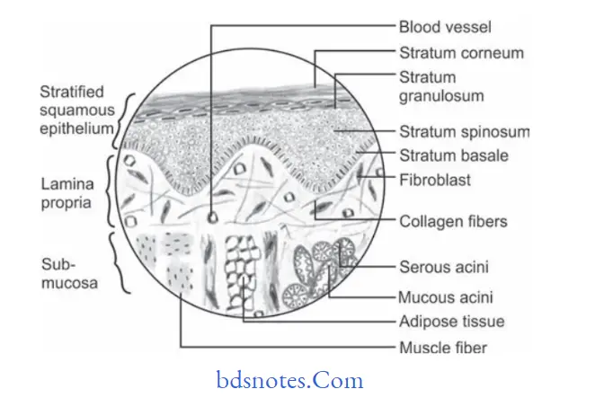
  • It is a special stain which is used for connective tissue elements. It is used for differentiating between connective tissue fiers and muscle fiers.
  • Epithelium (cell and cytoplasm): It takes greenish-yellow stain.
  • Collagen fiers are red
  • Muscle fiers are yellow
  • Nuclei of cells are blue black.

“Importance of studying special stains for better diagnostic outcomes: Questions explained”

Mallory stain

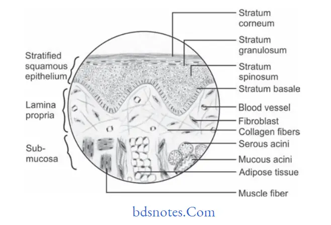
  • It is a special stain for keratin that stains deep orange. It is used in hyperkeratotic lesions.
  • The epithelium is royal blue.
  • Collagen fiers are royal blue.
  • Muscle fiers are royal blue.
  • Keratin layers are orange.
  • Nucleus is blue black.

Benign and malignant tumors of Oral cavity Mallory stain

“Common challenges in applying special stains effectively: FAQs provided”

Periodic Acid Schiff’s Stain Or Pas Stain

Benign and malignant tumors of Oral cavity PAS stain

“Steps to explain types of special stains: H&E vs PAS: Q&A guide”

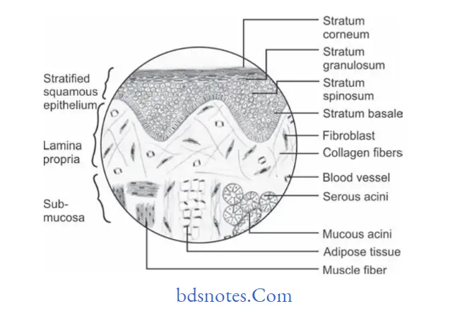
It is a special stain for mucopolysaccharide granules.

These are prominently seen in basement membrane, intercellular spaces and keratin layer.

It is used to detect continuity of basement membrane in intra­ epithelial carcinoma and squamous cell carcinoma.

  • Epithelium and connective tissue is pink.
  • Collagen fiers are pink.
  • Muscle fiers are pink.
  • Nucleus is blue black.
  • Granules are of magenta color.

“Role of hematoxylin and eosin (H&E) in histopathology: Questions answered”

Masson’s trichrome stain

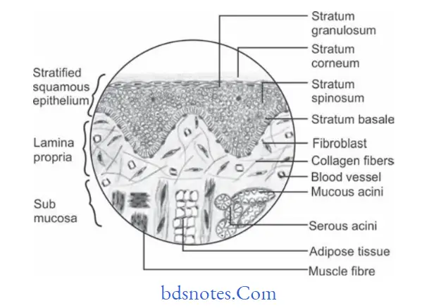
It is a special stain used to diffrentiate between collagen fiers and muscle fiers. It demonstrates connective tissue disorders like leiomyosarcoma and rhabdomyosarcoma.

  • Epithelium is red.
  • Muscle fiers are bluish violet.
  • Collagen fiers and blood vessels are blue.

“Early warning signs of issues addressed by understanding special stain types: Common questions”

Benign and malignant tumors of Oral cavity Masson’s trichrome stain

Filed Under: Oral Pathology

Reader Interactions

Leave a Reply Cancel reply

Your email address will not be published. Required fields are marked *

Primary Sidebar

Recent Posts

  • Branchial Cleft Cyst: Background, Pathophysiology, Etiology
  • Maxillary Nerve: Origin, Course, And Branches
  • The Father Of Anatomy And A Great Anatomist Herophilus
  • Bone Structure – Anatomy
  • The External Carotid Artery: Anatomy, Branches, And Functions
  • Occipitofrontalis Muscle
  • Superficial Temporal Artery
  • Platysma Muscle
  • Cartilage
  • Cauda Equina And Conus Medullaris Syndromes
  • Subcutaneous Injections And Device Management
  • Types Of Circulation: Pulmonary, Systemic, And Portal
  • Structure Of Skeletal Muscle
  • Elastic Cartilage
  • Cellular Organelles And Structure
  • The Golgi Apparatus – The Cell
  • The Cytoplasmic Inclusions Of Certain Plant Cells
  • Dental Abscess
  • Laser Surgery
  • Our Facial Muscles And Their Functions

Copyright © 2026 · Magazine Pro on Genesis Framework · WordPress · Log in