• Skip to main content
  • Skip to secondary menu
  • Skip to primary sidebar
  • About Us
  • Terms of Use
  • Privacy Policy
  • Disclaimer
  • Contact Us
  • Sitemap

BDS Notes

BDS notes, Question and Answers

  • Public Health Dentistry
  • Periodontics
  • Pharmacology
  • Pathology
  • Orthodontics
    • Anchorage In Orthodontics
    • Mandibular Growth, Functional Matrix
    • Retention and Relapse
  • General Surgery
    • Cysts: Types, Causes, Symptoms
    • Maxillofacial Fractures, Disorders, and Treatments
    • Lymphatic Disorders
    • Neurological and Facial Disorders
  • Temporal And Infratemporal Regions
    • Spinal and Neuroanatomy
  • Dental Materials
    • Dental Amalgam
Home » Histopathology of Moderately Differentiated Squamous Cell Carcinoma: Key Features Explained

Histopathology of Moderately Differentiated Squamous Cell Carcinoma: Key Features Explained

July 22, 2025 by Joankessler parkland Leave a Comment

Histopathology of Moderately Differentiated Squamous Cell Carcinoma: Key Features Explained

Question. Describe the histopathology of moderately differentiated squamous cell carcinoma. 

Answer.

“Common challenges in identifying moderately differentiated SCC effectively: FAQs provided”

Moderately Differentiated Squamous Cell Carcinoma Histopathology

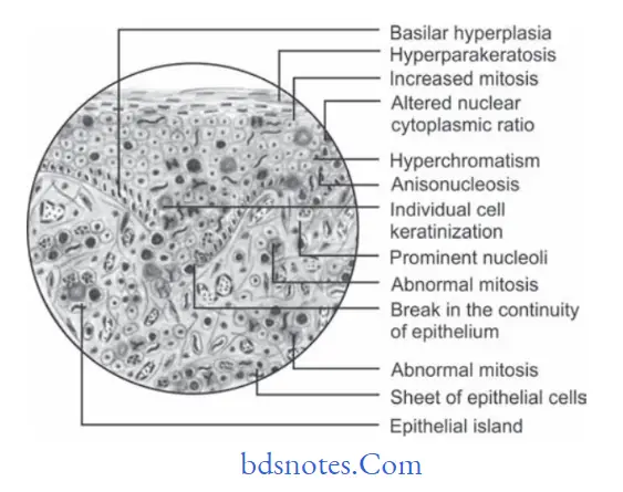
  • In it the tumor cells are usually more severely dysplastic than of well well-differentiated type.
  • Malignant epithelial cells produce little or no keratin and they exhibit greater number of mitotic division.
  • Formation of epithelial islands is diminished since tumor cells do not mature properly.
  • Malignant tumor cells are recognized as stratifid squamous epithelial cells.

“Understanding the role of histopathology in diagnosing squamous cell carcinoma: Q&A explained”

Benign and malignant tumors of Oral cavity Moderately differentiated squamous cell carcinoma

“Importance of studying histopathology of squamous cell carcinoma: Questions explained”

squamous cell carcinoma histopathology


Filed Under: Oral Pathology

Reader Interactions

Leave a Reply Cancel reply

Your email address will not be published. Required fields are marked *

Primary Sidebar

Recent Posts

  • Branchial Cleft Cyst: Background, Pathophysiology, Etiology
  • Maxillary Nerve: Origin, Course, And Branches
  • The Father Of Anatomy And A Great Anatomist Herophilus
  • Bone Structure – Anatomy
  • The External Carotid Artery: Anatomy, Branches, And Functions
  • Occipitofrontalis Muscle
  • Superficial Temporal Artery
  • Platysma Muscle
  • Cartilage
  • Cauda Equina And Conus Medullaris Syndromes
  • Subcutaneous Injections And Device Management
  • Types Of Circulation: Pulmonary, Systemic, And Portal
  • Structure Of Skeletal Muscle
  • Elastic Cartilage
  • Cellular Organelles And Structure
  • The Golgi Apparatus – The Cell
  • The Cytoplasmic Inclusions Of Certain Plant Cells
  • Dental Abscess
  • Laser Surgery
  • Our Facial Muscles And Their Functions

Copyright © 2026 · Magazine Pro on Genesis Framework · WordPress · Log in