Enamel Hypoplasia
Question: Write a short note on enamel hypoplasia.
Answer.
Enamel Hypoplasia
It is defined as an incomplete or defective formation of the organic enamel matrix.
Enamel hypoplasia is of two types, i.e.
- Acquired enamel hypoplasia.
- Environmental enamel hypoplasia.
Enamel hypoplasia diagnosis
Hypoplasia Acquired Enamel
Factors Associated with Acquired Enamel Hypoplasia
Two types of factors are associated with acquired enamel hypoplasia:
- Hypoplasia Local factors:
- Local factors are infection, trauma, and radiotherapy
- Idiopathic factors.
- When local infection or trauma causes damage to ameloblast cells during odontogenesis, it may lead to a defect in enamel formation in an isolated permanent tooth. This is known as focal enamel hypoplasia.
- Focal enamel hypoplasia is caused by to periapical spread of infection from a carious deciduous tooth or trauma to the deciduous tooth; the permanent tooth bud affected in this process is known as Turner’s tooth.
2. Environmental or systemic factors:
Systemic or environmental disturbances in the functioning of ameloblasts at a specific period during odontogenesis of teeth manifest as a horizontal line of small pits or grooves on enamel surfaces. This line on the tooth surface indicates a zone of enamel hypoplasia and corresponds to the time of development and duration of the insult.
The following are the various factors that lead to systemic or environmental disturbances:
Turner’s tooth
Prenatal period: The prenatal infections are rubella, syphilis.
- There is a presence of internal disease
- There are excess fluoride ions.
Hypoplasia Neonatal period: During this period, enamel hypoplasia is caused due to:
- Hemolytic disease of the newborn
- Birth injury
- Premature delivery
- Prolong labor
- Low birth weight.
Hypoplasia Postnatal period: During this period, enamel hypoplasia is due to:
- Severe childhood infection
- Prolonged fever due to infectious disease in childhood
- Nutritional deficiency
- Hypocalcemia
- Rickets
- Celiac disease.
Enamel hypoplasia symptoms
Environmental Enamel Hypoplasia
In it, either primary or permanent dentition is involved.
Both enamel and dentin are affected.
Enamel Hypoplasia Causes
- Nutritional deficiency
- Exanthematous diseases
- Congenital syphilis
- Hypocalcemia
- Birth injury, prematurity, Rh-incompatibility
- Local infection/trauma
- Ingestion of chemicals.
Enamel hypoplasia in children
Enamel Hypoplasia Types
- Mild: Few small grooves, pits, or fissures in the enamel surface.
- Moderate: Deep pits are arranged horizontally across the tooth.
- Severe: A Considerable portion of the enamel may be lost.
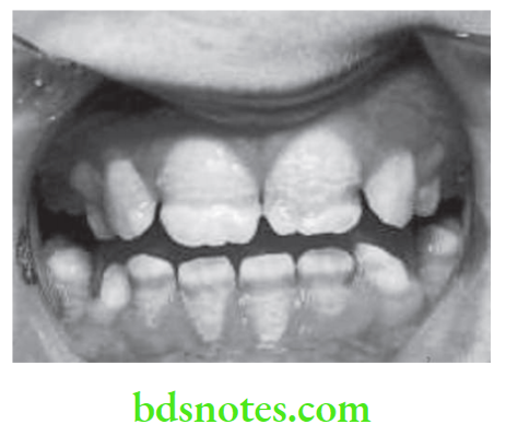
Enamel Hypoplasia Clinical Features
- Usually pitting type of hypoplasia is seen in deficiency of vitamin A, C, and during tooth formation.
- In case of congenital syphilis, incisors show Hutchinson’s teeth characterized by screw driven-shaped incisors and mulberry molars characterized by irregular enamel on the occlusal surface of the crown.
- Presence of Turner’s teeth in local infection.
Acquired enamel hypoplasia
Enamel Hypoplasia Management
- Teeth that are affected by enamel hypoplasia are more susceptible to dental caries as compared to normal teeth. Restoration should be confined to the area of involvement.
- In severe enamel hypoplasia chrome steel crown is given. Severe forms need composite restoration or a full ceramic crown.
- If sensitivity is present, desensitizing paste is prescribed.
- In mild hypoplastic molars, pits and fissures are placed over the occlusal surface, and 6 6-month re-evaluation is done.
- In patients with more demineralization, affected enamel should be removed, and the tooth should be restored with composite.
- Reduction of tooth and crown placement can also be done.
- In severe cases, extraction can be done.
- In anterior teeth, porcelain veneers or zirconia crowns are indicated.
Leave a Reply