How to Identify and Diagnose Basal Cell Carcinoma Early
Question 1. Write a note on precancerous lesions.
Answer:
Precancerous lesions
Precancerous lesions are defined as morphologically altered tissue in which cancer is most likely to occur than in its apparently normal counterpart.
- Leukoplakia
- Erythroplakia
- Mucosal changes associated with smoking habits
- Carcinoma in situ
- Bowen disease
- Actinic keratosis, cheilitis, and elastosis.
Basal cell carcinoma
“Understanding basal cell carcinoma: Causes and symptoms”
Question 2. Write a short note on basal cell carcinoma.
Answer:
Basal cell carcinoma
Basal cell carcinoma is a common locally aggressive non-metastatizing malignant neoplasm of skin that is composed of a medullary pattern of basaloid cells.
Basal cell carcinoma Clinical Features
- Basal cell carcinoma develops mostly in middle-aged people, preferably in the 4th decade of life.
- Males are more commonly affected than females.
- The neoplasm commonly occurs over the hair-bearing areas of facial skin. The orofacial areas particularly vulnerable to these lesions are the upper lip, nasolabial folds, periorbital region, cheek, forehead, ear, etc.
- The neoplasm initiates as a slow-growing, firm, slightly elevated, small nodule.
- It gradually enlarges and develops a central crusted ulcer with an elevated, smooth, rolled border.
- There may be intermittent bleeding from the ulcer.
Early signs of basal cell carcinoma
“Complications of untreated basal cell carcinoma”
“Importance of early detection of basal cell carcinoma”
Basal cell carcinoma Gross Features
- Grossly the most common pattern is noduloulcerative basal cell carcinoma in which a slow-growing small nodule undergoes central ulceration with pearly, rolled margins.
A tumor enlarges in size by burrowing and destroying the tissues locally like a rodent and so it is named as rodent ulcer. - Less frequently non-ulcerated nodular patterns, pigmented basal cell carcinoma, and firosingvariants are encountered.
“Treatment options for early-stage basal cell carcinoma”
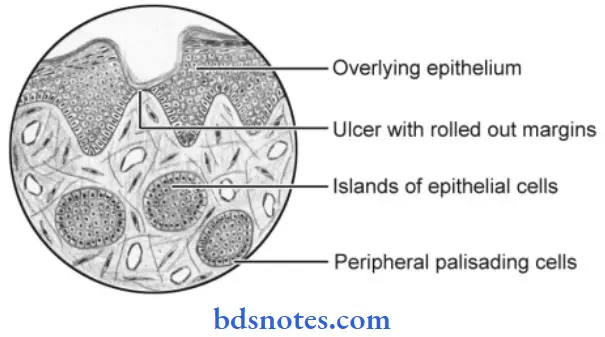
Basal cell carcinoma Histopathology
- Histologically, basal cell carcinoma is characterized by neoplastic proliferation of basaloid epithelial cells in the form of multiple solid islands or strands.
- These cells arise from the basal cell layer of the epidermis and they invade into the underlying dermis.
- The cells in the periphery of the tumor islands are columnar in shape and they often resemble the basal layer of the oral epithelium with hyperchromatic nuclei.
- These tumor cells do not show any feature of abnormal mitosis.
How to identify basal cell carcinoma
“Role of surgical excision in treating basal cell carcinoma”
- The cells are uniform in shape and size and in their staining reaction. Moreover, these cells often have a palisaded arrangement.
- The central cells of the tumor islands may be polyhedral, oval, round, or even spindle-shaped.
- The fibrous connective tissue stroma reveals varying degrees of cellularity and it contains a large number of elastic fibers.
Leave a Reply