Liquefaction Necrosis
Question 1. Write A Short Note On Liquefaction Necrosis.
Answer:
It is also known as colliquative necrosis
- When necrosed cells and tissues are converted into structure-less flids, it is called liquefactive necrosis.
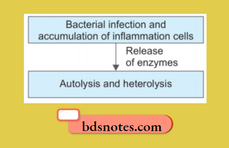
- It occurs commonly due to ischemia and bacterial or fungal infection due to the degradation of tissues by the action of powerful hydrolytic enzymes.
- The cellular structure of the organ is lost, and the tissue is digested, it is converted into a liquefied mass, which appears creamy yellow and is called pus.
- The common examples are infarct brain and abscess cavity.
Liquefaction necrosis
Liquefaction Necrosis Mechanism
Liquefaction Necrosis Gross Features
- Nerve tissues become liquefied after necrosis because they contain more lipids and water, which do not coagulate.
- The affected area is set with a liquefied center containing necrotic debris.
Liquefaction Necrosis Microscopic Features
- Microscopically liquefied tissue has no structure. The cystic space contains necrotic debris, phagocytosed material, and macrophages.
- The cyst wall is formed by the proliferation of capillaries, inflammatory cells, and glial cells in the case of the brain and proliferating fibroblasts in the case of the abscess cavity.
Question 2. Write A short note on hemosiderin.
Answer:
It is a hemoprotein-derived pigment.
- It is formed by the aggregates of ferritin and is identified in light microscopy.
- It is a golden yellow to brown granular pigment, especially within mononuclear phagocytes of bone marrow, spleen, and liver.
- Hemosiderin is a ferric iron, which is demonstrated by the Prussian blue reaction.
- Excessive storage of hemosiderin occurs in a situation where there is an increased breakdown of red cells or systemic overload of iron due to primary hemochromatosis and secondary causes such as thalassemia, sideroblastic anemia, and alcoholic cirrhosis.
- Effects of hemosiderin excess are:
- Localized: It develops due to hemorrhage in local tissues. With the lysis of red cells, hemoglobin is liberated and is taken up by macrophages and is degraded and stored as hemosiderin, For example,e. Hemorrhage gee in tissues, black eye, infarction, brown induration of lung.
- Systemic: Systemic overload of iron leads to generalized hemosiderosis. It is of two types:
- Parenchymal deposition in the liver, pancreas, kidney, heart, and skin.
- Reticuloendothelial cell deposits in the liver, spleen, and bone marrow
Difference between coagulative and liquefactive necrosis
Following are the examples of systemic hemosiderosis:
- Acquired hemosiderosis in chronic hemolytic disorders, blood transfusion, parenteral administration of iron.
- Hereditary hemochromatosis
- Excessive dietary intake, i.e., in Bantu’s disease.
Leave a Reply