• Skip to main content
  • Skip to secondary menu
  • Skip to primary sidebar
  • About Us
  • Terms of Use
  • Privacy Policy
  • Disclaimer
  • Contact Us
  • Sitemap

BDS Notes

BDS notes, Question and Answers

  • Public Health Dentistry
  • Periodontics
  • Pharmacology
  • Pathology
  • Orthodontics
    • Anchorage In Orthodontics
    • Mandibular Growth, Functional Matrix
    • Retention and Relapse
  • General Surgery
    • Cysts: Types, Causes, Symptoms
    • Maxillofacial Fractures, Disorders, and Treatments
    • Lymphatic Disorders
    • Neurological and Facial Disorders
  • Temporal And Infratemporal Regions
    • Spinal and Neuroanatomy
  • Dental Materials
    • Dental Amalgam
Home » Anatomy Of The Temporomandibular Joint

Anatomy Of The Temporomandibular Joint

July 28, 2025 by Kristensmith Taylor Leave a Comment

Anatomy Of The Temporomandibular Joint

Enumerate the ligaments and functions of TMJ.
Answer:

Ligaments:

1. Fibrous capsule:

  • Attached above to the articular tubercle, mandibular fossa, and below to the neck of the mandible.

2. Lateral/temporomandibular ligament:

  • Attached above to the articular tubercle.
  • Below to the posterolateral aspect of the neck of the mandible.
  • It reinforces and strengthens the capsular ligament.

“Understanding the TMJ through FAQs: Structure, functions, and uses explained”

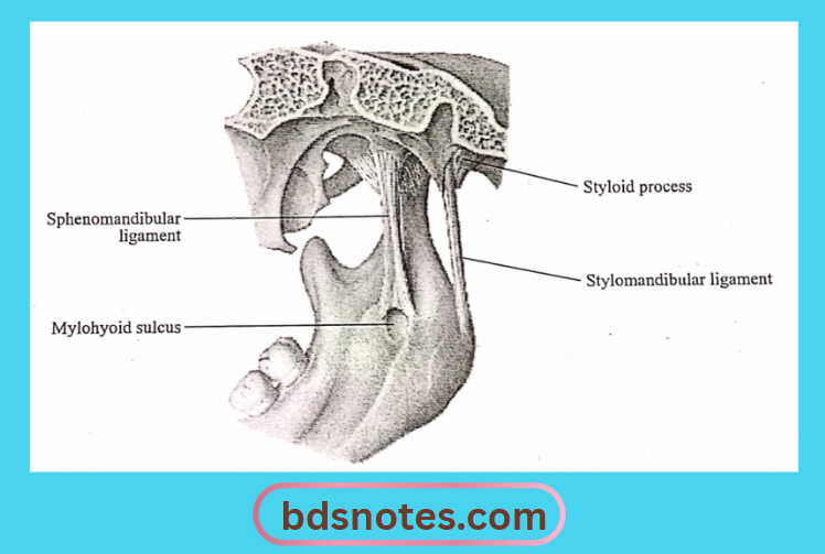
3. Sphenomandibular ligament:

  • It is an accessory ligament.
  • It arises from the spine of the sphenoid and from the petrotympanic fissures and ends at the lingula of the mandible.
  • It is a remnant of Meckel’s cartilage.

4. Stylomandibular ligament:

  • It is also an accessory ligament.
  • It is attached above the lateral surface of the styloid process and below to the angle and ramus of the mandible.

Capsular Anatomy Of Temporomandibular Joint

“Importance of studying TMJ anatomy for dental students: Questions explained”

Mylohyoid Sulcus And Ligament Relations

“Common challenges in mastering TMJ anatomy notes effectively: FAQs provided”

5. Otomandibular ligaments:

  • These are discomalleolar and tympano mandibular ligaments.
  • They connect callers to the TMJ disk and to the sphenomandibular ligaments.

Temporomandibular Joints Functions:

1. Protraction/forward movement of the mandible.

  • During this movement the articular disc of the TMJ glides forward over the upper articular surface, the head of the mandible moving with it.

2. Retraction of mandible:

  • During this, the articular disc glides backward over the upper articular surface.

“Factors influencing success with TMJ studies: Q&A”

3. Slight opening of the mouth.

  • The head of the mandible moves on the undersurface of the disc.

4. Wide opening of the mouth.

  • It is followed by protraction.

5. Chewing movements/side-to-side movements of the mandible.

Filed Under: Anatomy

Reader Interactions

Leave a Reply Cancel reply

Your email address will not be published. Required fields are marked *

Primary Sidebar

Recent Posts

  • Branchial Cleft Cyst: Background, Pathophysiology, Etiology
  • Maxillary Nerve: Origin, Course, And Branches
  • The Father Of Anatomy And A Great Anatomist Herophilus
  • Bone Structure – Anatomy
  • The External Carotid Artery: Anatomy, Branches, And Functions
  • Occipitofrontalis Muscle
  • Superficial Temporal Artery
  • Platysma Muscle
  • Cartilage
  • Cauda Equina And Conus Medullaris Syndromes
  • Subcutaneous Injections And Device Management
  • Types Of Circulation: Pulmonary, Systemic, And Portal
  • Structure Of Skeletal Muscle
  • Elastic Cartilage
  • Cellular Organelles And Structure
  • The Golgi Apparatus – The Cell
  • The Cytoplasmic Inclusions Of Certain Plant Cells
  • Dental Abscess
  • Laser Surgery
  • Our Facial Muscles And Their Functions

Copyright © 2026 · Magazine Pro on Genesis Framework · WordPress · Log in