• Skip to main content
  • Skip to secondary menu
  • Skip to primary sidebar
  • About Us
  • Terms of Use
  • Privacy Policy
  • Disclaimer
  • Contact Us
  • Sitemap

BDS Notes

BDS notes, Question and Answers

  • Public Health Dentistry
  • Periodontics
  • Pharmacology
  • Pathology
  • Orthodontics
    • Anchorage In Orthodontics
    • Mandibular Growth, Functional Matrix
    • Retention and Relapse
  • General Surgery
    • Cysts: Types, Causes, Symptoms
    • Maxillofacial Fractures, Disorders, and Treatments
    • Lymphatic Disorders
    • Neurological and Facial Disorders
  • Temporal And Infratemporal Regions
    • Spinal and Neuroanatomy
  • Dental Materials
    • Dental Amalgam
Home » A Complete Guide To The Primary Mandibular Second Molar

A Complete Guide To The Primary Mandibular Second Molar

February 5, 2026 by Kristensmith Taylor Leave a Comment

A Complete Guide To The Primary Mandibular Second Molar

Describe in detail about Primary Mandibular Second Molar.
Answer:

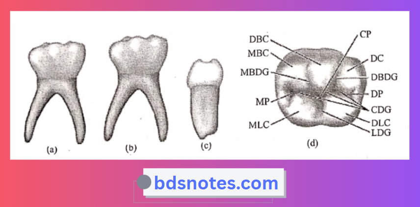
Primary Mandibular Second Molar Buccal aspect:

  • It is wider at the cervical portion.
  • Mesiobuccal and distobuccal developmental grooves divide the buccal surface into mesiobuccal, buccal, and distobuccal cusp.
  • Roots appear slender and long.
  • Roots flare mesiodistally at the middle and apical third.
  • The point of bifurcation of the roots is just below the CEJ.

Primary Mandibular Second Molar Lingual aspect:

  • Two equal cusps with a lingual groove between them are seen.
  • The cervical line is relatively straight.
  • A portion of each of the three buccal cusps may be seen. Roots appear slender and long.

Primary Mandibular Second Molar Mesial aspects:

  • The tooth seems constricted occlusal. The lingual cusp is longer.
  • The cervical line is regular.
  • The mesial root is broad and flat.

Primary Mandibular Second Molar Distal aspect:

  • Mesiobuccal and distobuccal cusps are seen distolingual cusp appears well developed.
  • The triangular ridge extends from the distolingual cusp tip to the distal marginal ridge.
  • The cervical line of the crown is regular.
  • The distal root is broad and flat and tapers more at the apical end.

Primary Mandibular Second Molar Occlusal aspect:

  • It is somewhat rectangular.
  • Buccal cusps are the same in size, while both lingual cusps are equal.
  • Well-defined triangular ridges are seen.
  • The occlusal surface shows.
  • Mesial triangular fossa.
  • Inside the mesial marginal ridge.
  • Distal triangular fossa.
  • Mesial to the distal marginal ridge.
  • Central developmental groove.
  • Extends from mesial triangular fossa to distal triangular fossa.
  • Two buccal grooves-mesial and distal.
  • Lingual developmental groove.
  • Supplemental grooves.
  • Seen over slopes of triangular ridges and in mesial and distal triangular fossae.
  • The mesial marginal ridge is well developed.
  • Crown converge distally.

Pri Mandibular 2nd Molar Occlusal Surface

Filed Under: Anatomy

Reader Interactions

Leave a Reply Cancel reply

Your email address will not be published. Required fields are marked *

Primary Sidebar

Recent Posts

  • Branchial Cleft Cyst: Background, Pathophysiology, Etiology
  • Maxillary Nerve: Origin, Course, And Branches
  • The Father Of Anatomy And A Great Anatomist Herophilus
  • Bone Structure – Anatomy
  • The External Carotid Artery: Anatomy, Branches, And Functions
  • Occipitofrontalis Muscle
  • Superficial Temporal Artery
  • Platysma Muscle
  • Cartilage
  • Cauda Equina And Conus Medullaris Syndromes
  • Subcutaneous Injections And Device Management
  • Types Of Circulation: Pulmonary, Systemic, And Portal
  • Structure Of Skeletal Muscle
  • Elastic Cartilage
  • Cellular Organelles And Structure
  • The Golgi Apparatus – The Cell
  • The Cytoplasmic Inclusions Of Certain Plant Cells
  • Dental Abscess
  • Laser Surgery
  • Our Facial Muscles And Their Functions

Copyright © 2026 · Magazine Pro on Genesis Framework · WordPress · Log in