• Skip to main content
  • Skip to secondary menu
  • Skip to primary sidebar
  • About Us
  • Terms of Use
  • Privacy Policy
  • Disclaimer
  • Contact Us
  • Sitemap

BDS Notes

BDS notes, Question and Answers

  • Public Health Dentistry
  • Periodontics
  • Pharmacology
  • Pathology
  • Orthodontics
    • Anchorage In Orthodontics
    • Mandibular Growth, Functional Matrix
    • Retention and Relapse
  • General Surgery
    • Cysts: Types, Causes, Symptoms
    • Maxillofacial Fractures, Disorders, and Treatments
    • Lymphatic Disorders
    • Neurological and Facial Disorders
  • Temporal And Infratemporal Regions
    • Spinal and Neuroanatomy
  • Dental Materials
    • Dental Amalgam
Home » Understanding The Morphology Of Primary Mandibular First Molars

Understanding The Morphology Of Primary Mandibular First Molars

February 5, 2026 by Kristensmith Taylor Leave a Comment

Understanding The Morphology Of Primary Mandibular First Molars

Describe in detail about Primary Mandibular First Molar.
Answer:

Primary Mandibular First Molar Buccal aspect:

  • It has a straight outline.
  • The Crown constricts at the cervix.
  • The distal portion of the crown is shorter than the mesial portion.
  • Cervical line dip apically.
  • The mesial cusp is larger than the distal cusp.
  • Development depression divides them. The roots are long and slender

Primary Mandibular First Molar Lingual aspect:

  • Crown and root converge lingually.
  • The distolingual cusp is rounded.
  • The mesiolingual cusp is long and sharp.
  • Developmental groove occurs between the distolingual and mesiolingual cusp.
  • The cervical line is straight.

Primary Mandibular First Molar Mesial aspect:

  • Mesiobuccal and mesiolingual cusps are seen.
  • The mesial marginal ridge is well-developed.
  • The cervical line slants upward.
  • The root end is flat and almost square.

Primary Mandibular First Molar Distal aspect:

  • The cervical line is almost straight.
  • The distal marginal ridge is not well defined.
  • The distal root is rounder, shorter, and tapers apically.

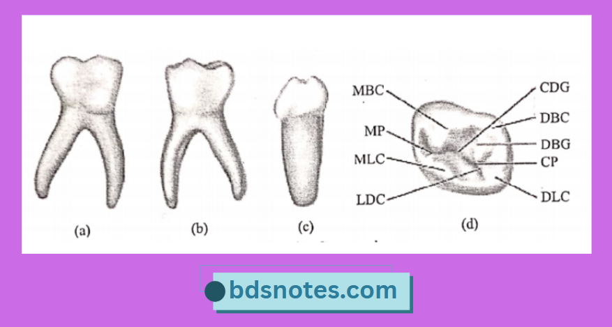
Primary Mandibular First Molar Occlusal aspect:

  • The mesiolingual cusp is the largest and most well-developed.
  • The buccal developmental groove divides the two buccal cusps.
  • The central developmental groove separates the mesiobuccal and mesiolingual cusps.
  • Two supplemental grooves are seen.
  • The mesiobuccal cusp exhibits a well-defined triangular ridge.
  • Lingual developmental grooves separate media lingual and distolingual cusp.

Primary Mandibular Molar Mbc Mp Mlc

Filed Under: Anatomy

Reader Interactions

Leave a Reply Cancel reply

Your email address will not be published. Required fields are marked *

Primary Sidebar

Recent Posts

  • Branchial Cleft Cyst: Background, Pathophysiology, Etiology
  • Maxillary Nerve: Origin, Course, And Branches
  • The Father Of Anatomy And A Great Anatomist Herophilus
  • Bone Structure – Anatomy
  • The External Carotid Artery: Anatomy, Branches, And Functions
  • Occipitofrontalis Muscle
  • Superficial Temporal Artery
  • Platysma Muscle
  • Cartilage
  • Cauda Equina And Conus Medullaris Syndromes
  • Subcutaneous Injections And Device Management
  • Types Of Circulation: Pulmonary, Systemic, And Portal
  • Hierarchical Organization Of Skeletal Muscle Tissue
  • Elastic Cartilage Histology Short Note For Medical Exams
  • Cellular Organelles And Structure
  • The Golgi Apparatus – The Cell
  • The Cytoplasmic Inclusions Of Certain Plant Cells
  • Dental Abscess
  • Laser Surgery
  • Our Facial Muscles And Their Functions

Copyright © 2026 · Magazine Pro on Genesis Framework · WordPress · Log in