• Skip to main content
  • Skip to secondary menu
  • Skip to primary sidebar
  • About Us
  • Terms of Use
  • Privacy Policy
  • Disclaimer
  • Contact Us
  • Sitemap

BDS Notes

BDS notes, Question and Answers

  • Public Health Dentistry
  • Periodontics
  • Pharmacology
  • Pathology
  • Orthodontics
    • Anchorage In Orthodontics
    • Mandibular Growth, Functional Matrix
    • Retention and Relapse
  • General Surgery
    • Cysts: Types, Causes, Symptoms
    • Maxillofacial Fractures, Disorders, and Treatments
    • Lymphatic Disorders
    • Neurological and Facial Disorders
  • Temporal And Infratemporal Regions
    • Spinal and Neuroanatomy
  • Dental Materials
    • Dental Amalgam
Home » Specialized Mucosa Of The Tongue: Structure And Features

Specialized Mucosa Of The Tongue: Structure And Features

February 6, 2026 by Kristensmith Taylor Leave a Comment

Specialized Mucosa Of The Tongue: Structure And Features

Question 1. Specialized mucosa
Answer:

  • The mucous membrane covering the dorsum of the tongue is called specialized mucosa

Dorsum of the tongue:

  • It is rough and irregular
  • It is divided by the V-shaped groove called sulcus terminals into
  • Anterior two-third or papillary part
  • Posterior one third or lymphatic part

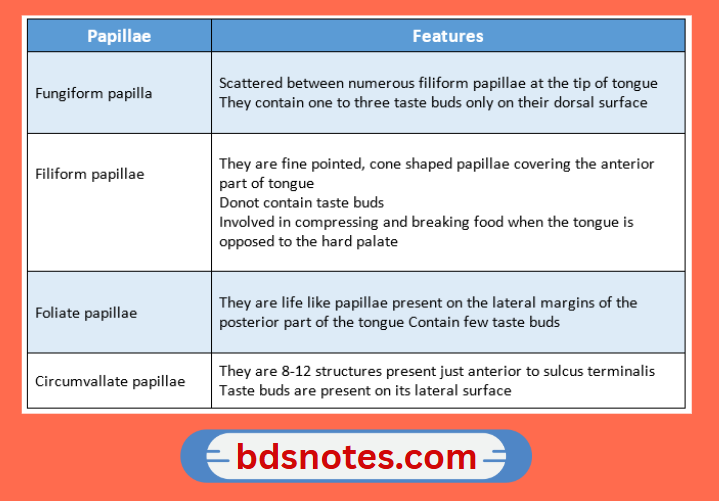
Papillae of the tongue:

Tongue Papillae Features


Posterior one-third of the tongue:

  • It contains round or oval prominences called lingual follicles
  • Each of these has one or more lymph nodules
  • The lingual follicles together form lingual tonsil

Question 2. Lining mucosa 
Answer:

  • The oral mucosa covering the underside of the tongue, inside of the lips, cheeks, floor of the mouth, vestibule, and alveolar mucosa are classified as lining mucosa.

Histology:

1. Epithelium

  • Non-keratinized stratified squamous epithelium
  • The surface is flexible to withstand stretching

2. Junction between epithelium and lamina propria

  • Smooth
  • Connective tissue papilla often penetrates into the epithelium

3. Lamina propria

  • Thicker
  • Contains fewer irregular collagen fibers and elastic fibers

4. Submucosa

  • The mucosa of the soft palate is separated from the loose and highly glandular submucosa by a layer of elastic fibers

Functions:

  • Collagen fibers help to stretch the mucosa to a certain limit
  • Elastic fibers tend to control the extensibility of the mucosa

Filed Under: Anatomy

Reader Interactions

Leave a Reply Cancel reply

Your email address will not be published. Required fields are marked *

Primary Sidebar

Recent Posts

  • Branchial Cleft Cyst: Background, Pathophysiology, Etiology
  • Maxillary Nerve: Origin, Course, And Branches
  • The Father Of Anatomy And A Great Anatomist Herophilus
  • Bone Structure – Anatomy
  • The External Carotid Artery: Anatomy, Branches, And Functions
  • Occipitofrontalis Muscle
  • Superficial Temporal Artery
  • Platysma Muscle
  • Cartilage
  • Cauda Equina And Conus Medullaris Syndromes
  • Subcutaneous Injections And Device Management
  • Types Of Circulation: Pulmonary, Systemic, And Portal
  • Structure Of Skeletal Muscle
  • Elastic Cartilage
  • Cellular Organelles And Structure
  • The Golgi Apparatus – The Cell
  • The Cytoplasmic Inclusions Of Certain Plant Cells
  • Dental Abscess
  • Laser Surgery
  • Our Facial Muscles And Their Functions

Copyright © 2026 · Magazine Pro on Genesis Framework · WordPress · Log in