• Skip to main content
  • Skip to secondary menu
  • Skip to primary sidebar
  • About Us
  • Terms of Use
  • Privacy Policy
  • Disclaimer
  • Contact Us
  • Sitemap

BDS Notes

BDS notes, Question and Answers

  • Public Health Dentistry
  • Periodontics
  • Pharmacology
  • Pathology
  • Orthodontics
    • Anchorage In Orthodontics
    • Mandibular Growth, Functional Matrix
    • Retention and Relapse
  • General Surgery
    • Cysts: Types, Causes, Symptoms
    • Maxillofacial Fractures, Disorders, and Treatments
    • Lymphatic Disorders
    • Neurological and Facial Disorders
  • Temporal And Infratemporal Regions
    • Spinal and Neuroanatomy
  • Dental Materials
    • Dental Amalgam
Home » Tooth-Gum Connection: Junctional Epithelium

Tooth-Gum Connection: Junctional Epithelium

February 6, 2026 by Kristensmith Taylor Leave a Comment

Tooth-Gum Connection: Junctional Epithelium

Write briefly the development of three stages of detinogingival junction.
Answer:

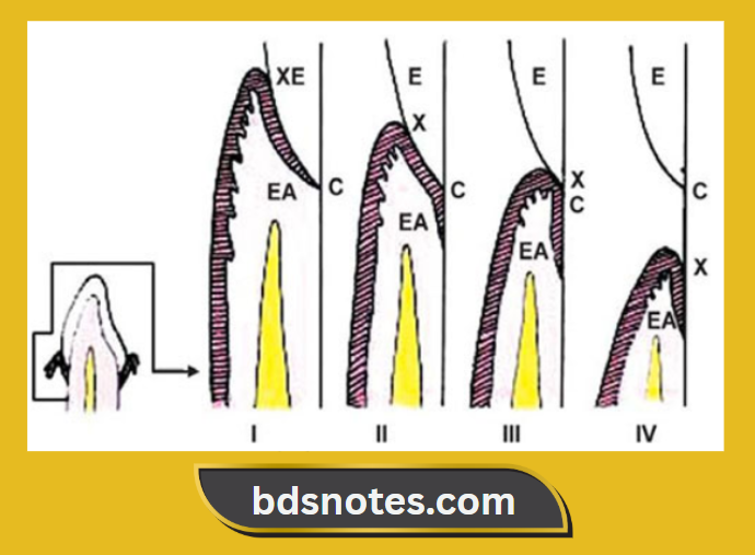
Stages of dentinogingival junction:

1. First stage physiologic:

  • Position of the bottom of the gingival sulcus. In the enamel-covered crown.
  • Position of the apical end of the attachment epithelium at tire cementoenamel junction.
  • The clinical crown is smaller than the anatomic crown.

Age:

  • Before 1 year of shedding of primary teeth.
  • In permanent teeth, at the age of 20 – 30 years.

2. Second stage – physiologic:

  • The bottom of the gingival sulcus is on the enamel.
  • The apical end of the attachment epithelium is on the cementum.
  • Fiber bundles present at the cervical parts of the cementum undergo dissolution.
  • This part later gets covered by the epithelium.
  • The apical shift of the gingival and transseptal fibers occurs.
  • Fibers are destroyed by the enzymes secreted by the epithelial cells, by plaque or by immunologic reactions.
  • The clinical crown is smaller than the anatomic crown.
  • Age: 40 years or later.

3. Third stage – pathologic:

  • The bottom of the gingival sulcus is at the cementoenamel junction.
  • Epithelium attachment is entirely on the cementum.
  • The tooth is exposed.
  • The epithelium shifts along the tooth surface.
  • The clinical crown is equal to the anatomic crown.

4. Fourth stage – pathologic:

  • It represents a gingival recession.
  • The entire attachment is on the cementum.
  • May occur even in the absence of periodontitis.
  • The clinical crown is longer than the atomic crown.

Age:

  • Varies
  • In some cases, occurs at the age of 20’s while absent even at the age of 50 or more.

Development Of Dentogingival Junction

Filed Under: Anatomy

Reader Interactions

Leave a Reply Cancel reply

Your email address will not be published. Required fields are marked *

Primary Sidebar

Recent Posts

  • Branchial Cleft Cyst: Background, Pathophysiology, Etiology
  • Maxillary Nerve: Origin, Course, And Branches
  • The Father Of Anatomy And A Great Anatomist Herophilus
  • Bone Structure – Anatomy
  • The External Carotid Artery: Anatomy, Branches, And Functions
  • Occipitofrontalis Muscle
  • Superficial Temporal Artery
  • Platysma Muscle
  • Cartilage
  • Cauda Equina And Conus Medullaris Syndromes
  • Subcutaneous Injections And Device Management
  • Types Of Circulation: Pulmonary, Systemic, And Portal
  • Hierarchical Organization Of Skeletal Muscle Tissue
  • Elastic Cartilage Histology Short Note For Medical Exams
  • Cellular Organelles And Structure
  • The Golgi Apparatus – The Cell
  • The Cytoplasmic Inclusions Of Certain Plant Cells
  • Dental Abscess
  • Laser Surgery
  • Our Facial Muscles And Their Functions

Copyright © 2026 · Magazine Pro on Genesis Framework · WordPress · Log in