Histology And Function Of The Dentogingival Junction In Oral Health
Write the development, structure, and different stages of the dentogingival junction.
Answer:
Dentogingival junction:
- It is the junction between the gingiva and the tooth.
- It represents a site of potential weakness in the continuous lining of the oral cavity.
Structure:
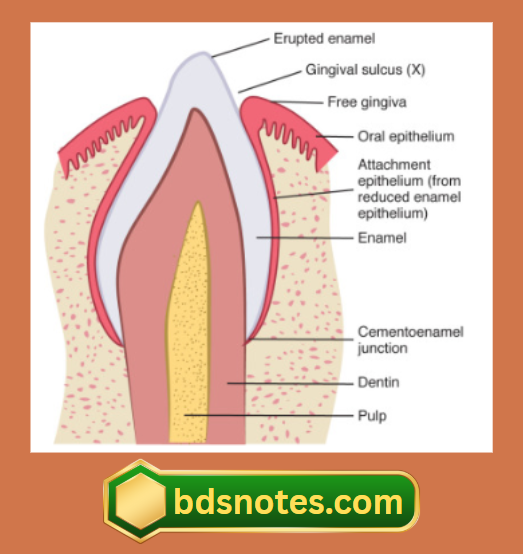
- The epithelium of the gingiva that attaches to the tooth is called junctional epithelium.
- The union between it and the tooth is called epithelial attachment.
- The junctional epithelium resembles reduced enamel epithelium.
- They have a basal layer and a few layers of flattened cells.
- It is non-differentiating, nonkeratinizing tissue.
- It is highly permeable with large intercellular spaces.
- Lymphocytes and plasma cells are seen in the connective tissue at the bottom of the gingival sulcus and below the attachment epithelium.
Development:
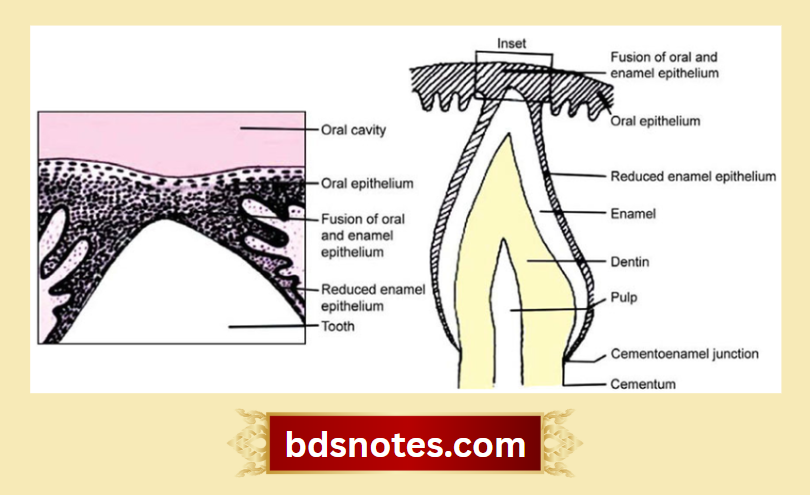
- After the formation of the enamel matrix, ameloblasts leave a thin membrane on the enamel surface, called the primary enamel cuticle.
- Then, the ameloblasts shorten and the epithelial enamel organ is reduced to a few layers of flat cuboidal cells called reduced enamel epithelium.
- It covers the entire enamel surface up to CEJ at the same time remains attached to the primary enamel cuticle.
- During the eruption, the tip of the tooth approaches the oral mucosa.
- By it, the reduced enamel epithelium and the oral epithelium meet and fuse.
- The remnants of the primary enamel cuticle are referred to as Nasmyth’s membrane.
- The epithelium covering the tip of the crown degenerates in its center through which it emerges into ora! cavity.
- Now, the reduced enamel epithelium is known as the primary attachment epithelium.
- At the margin of the gingival, the attachment epithelium becomes continuous with the oral epithelium.
- The reduced enamel epithelium gradually shortens due to which a shallow groove called gingival sulcus develops between the gingiva and the tooth surface.
- When the reduced enamel epithelium gets separated from the erupted tooth, the gingival sulcus further deepens.

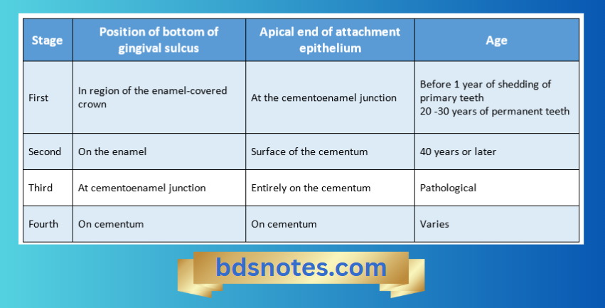
- During the first and second stages, a clinical crown is smaller than the anatomic crown.
- During the third stage, a clinical crown is equal to an anatomic crown.
- During the fourth stage, the clinical crown is larger than the anatomic crown.
Leave a Reply