Lining Mucosa: Histology And Its Role In Oral Surgery
Describe the lining mucosa.
Answer:
- The oral mucosa covering the underside of the tongue, inside of the lips, cheeks, floor of the mouth vestibule, and alveolar mucosa are classified as lining mucosa.
- The mucous membrane is movably attached to the deep structures and does not restrict the movement of lips, cheeks, and tongue.
- Where the lining mucosa covers muscle, the mucosa is fixed to the fascia.
Lining Mucosa Clinical considerations:
- Surgical incisions require sutures for closure.
- Injections are easy due to the ready dispersion of fluids.
- Infections spread rapidly.
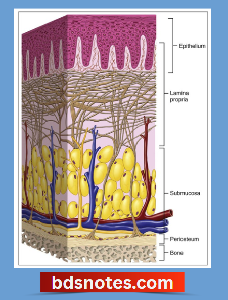
Lining Mucosa Histology:
1. Epithelium.
- Thick (400 pm)
- Non-keratinized stratified squamous epithelium.
- The surface is flexible to withstand stretching.
2. Junction between epithelium and lamina propria.
- Smooth
- Slender connective tissue papillae often penetrate into the epithelium.
3. Lamina propria.
- Thicker
- Contains fewer irregular collagen fibers.
- This helps the mucosa to be stretched to a certain extent.
- Also contains elastic fibers to control the extensibility of the mucosa.
4. Submucosa.
- The mucosa of the soft palate is separated from the loose and highly glandular submucosa by a layer of elastic fibers.
Attachments:
1. Lining mucosa covering the muscle.
- Attached by a mixture of collagen and elastic fibers
2. the alveolar mucosa and mucosa covering the floor of the mouth.
- Attached loosely to the underlying structures by a thick submucosa.
3. Mucosa of the underside of the tongue.
- Bound firmly to the underlying muscle.
Lining Mucosa Functions:
1. Collagen fibers.
- Helps to stretch the mucosa to a certain limit.
2. Elastic fibers.
- Tend to control the extensibility of the mucosa.
- During mastication.
- Retract the mucosa toward the muscle.
- Prevent it from bulging between the teeth and being bitten.
- Tend to restore the mucosa to its resting position after detention.
Leave a Reply