• Skip to main content
  • Skip to secondary menu
  • Skip to primary sidebar
  • About Us
  • Terms of Use
  • Privacy Policy
  • Disclaimer
  • Contact Us
  • Sitemap

BDS Notes

BDS notes, Question and Answers

  • Public Health Dentistry
  • Periodontics
  • Pharmacology
  • Pathology
  • Orthodontics
    • Anchorage In Orthodontics
    • Mandibular Growth, Functional Matrix
    • Retention and Relapse
  • General Surgery
    • Cysts: Types, Causes, Symptoms
    • Maxillofacial Fractures, Disorders, and Treatments
    • Lymphatic Disorders
    • Neurological and Facial Disorders
  • Temporal And Infratemporal Regions
    • Spinal and Neuroanatomy
  • Dental Materials
    • Dental Amalgam
Home » Maxillary Air Sinus: Size, Shape, and Anatomical Landmarks

Maxillary Air Sinus: Size, Shape, and Anatomical Landmarks

February 6, 2026 by Kristensmith Taylor Leave a Comment

Maxillary Air Sinus: Size, Shape, and Anatomical Landmarks

Describe Maxillary Air Sinus & its relations
Answer:

Maxillary Sinus:

  • It is largest of all sinuses
  • It lies in the body of maxilla

Maxillary Air Sinus Size:

  • Height-3.5 cm
  • Width-2.5 cm
  • Anteroposterior depth- 3.5 cm

Maxillary Air Sinus Boundaries:

  • It is pyramidal in shape
  • Base-directed medially towards lateral wall of the nose
  • Apex-Directed laterally in the zygomatic process of the maxilla
  • Roof- formed by Floor of the orbit
  • Floor- Formed by alveolar process of maxilla

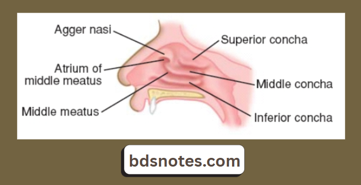
Maxillary Air Sinus Opening:

  • It opens into middle meatus of nose in the lower part of the hiatus semilunaris
  • A second opening is present at the posterior end of hiatus
  • This opening is reduced in intact skull as it is overlapped by the following.
    1. Uncinate process of the ethamoid- from above
    2. Ethamoidal process of inferior nasal concha- from below
    3. Perpendicular plate of palatine bone- from behind
    4. Descending process of the lacrimal bone- from front

Maxillary Air Sinus Relations:

Maxillary Sinus Relations

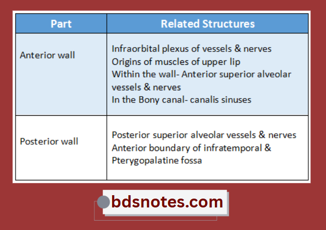
Maxillary Air Sinus Blood Supply:

Arterial Supply:

  • Facial artery
  • Infraorbital artery
  • Greater palatine artery

Maxillary Air Sinus Venous Drainage:

  • Facial vein
  • Pterygoid plexus of veins

Maxillary Air Sinus Nerve Supply:

  • Infraorbital nerve
  • Anterior, middle & posterior superior alveolar nerve

Maxillary Air Sinus Lymphatic Drainage:

  • Into Submandibular lymph nodes

Nasal Cavity Conchae And Meatus

Filed Under: Anatomy

Reader Interactions

Leave a Reply Cancel reply

Your email address will not be published. Required fields are marked *

Primary Sidebar

Recent Posts

  • Branchial Cleft Cyst: Background, Pathophysiology, Etiology
  • Maxillary Nerve: Origin, Course, And Branches
  • The Father Of Anatomy And A Great Anatomist Herophilus
  • Bone Structure – Anatomy
  • The External Carotid Artery: Anatomy, Branches, And Functions
  • Occipitofrontalis Muscle
  • Superficial Temporal Artery
  • Platysma Muscle
  • Cartilage
  • Cauda Equina And Conus Medullaris Syndromes
  • Subcutaneous Injections And Device Management
  • Types Of Circulation: Pulmonary, Systemic, And Portal
  • Structure Of Skeletal Muscle
  • Elastic Cartilage
  • Cellular Organelles And Structure
  • The Golgi Apparatus – The Cell
  • The Cytoplasmic Inclusions Of Certain Plant Cells
  • Dental Abscess
  • Laser Surgery
  • Our Facial Muscles And Their Functions

Copyright © 2026 · Magazine Pro on Genesis Framework · WordPress · Log in