• Skip to main content
  • Skip to secondary menu
  • Skip to primary sidebar
  • About Us
  • Terms of Use
  • Privacy Policy
  • Disclaimer
  • Contact Us
  • Sitemap

BDS Notes

BDS notes, Question and Answers

  • Public Health Dentistry
  • Periodontics
  • Pharmacology
  • Pathology
  • Orthodontics
    • Anchorage In Orthodontics
    • Mandibular Growth, Functional Matrix
    • Retention and Relapse
  • General Surgery
    • Cysts: Types, Causes, Symptoms
    • Maxillofacial Fractures, Disorders, and Treatments
    • Lymphatic Disorders
    • Neurological and Facial Disorders
  • Temporal And Infratemporal Regions
    • Spinal and Neuroanatomy
  • Dental Materials
    • Dental Amalgam
Home » Palatine Tonsils: Position, Relations, And Blood Supply Explained

Palatine Tonsils: Position, Relations, And Blood Supply Explained

February 6, 2026 by Kristensmith Taylor Leave a Comment

Palatine Tonsils: Position, Relations, And Blood Supply Explained

Question 1. Palatine tonsil
Answer:

Palatine tonsil Position:

Each tonsil occupies the triangular tonsillar sinus or fossa between palatoglossus and palatopharyngeal arches

Palatine tonsil Relations:

Palatopharyngeal Arch Tonsil Relation

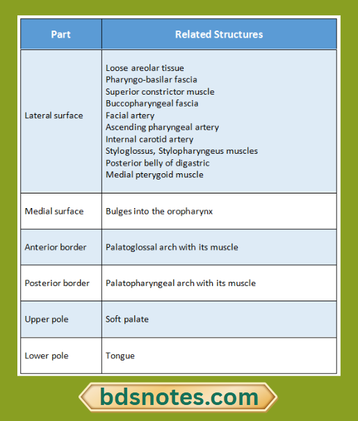
Palatine tonsil Blood supply:

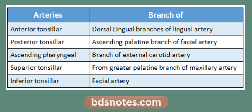
Palatine tonsil Arterial supply:

Palatine Tonsil Arterial Supply Diagram

Palatine tonsil Venous drainage:

  • Drains into pharyngeal venous plexus.

Question 2. Superior constrictor of pharynx
Answer:

Superior constrictor of pharynx Origin:

  • From the lower part of posterior border of medial pterygoid plate
  • From the pterygoid hamulus
  • From the posterior border of pterygomandibular raphe
  • From posterior end of the mylohyoid line of mandible
  • From the side of the tongue

Superior constrictor of pharynx Insertion:

  • Inserted into the median raphe on the posterior wall of the pharynx.

Superior constrictor of pharynx Nerve supply:

  • It is supplied by the cranial part of accessory nerve through the pharyngeal plexus

Filed Under: Anatomy

Reader Interactions

Leave a Reply Cancel reply

Your email address will not be published. Required fields are marked *

Primary Sidebar

Recent Posts

  • Branchial Cleft Cyst: Background, Pathophysiology, Etiology
  • Maxillary Nerve: Origin, Course, And Branches
  • The Father Of Anatomy And A Great Anatomist Herophilus
  • Bone Structure – Anatomy
  • The External Carotid Artery: Anatomy, Branches, And Functions
  • Occipitofrontalis Muscle
  • Superficial Temporal Artery
  • Platysma Muscle
  • Cartilage
  • Cauda Equina And Conus Medullaris Syndromes
  • Subcutaneous Injections And Device Management
  • Types Of Circulation: Pulmonary, Systemic, And Portal
  • Structure Of Skeletal Muscle
  • Elastic Cartilage
  • Cellular Organelles And Structure
  • The Golgi Apparatus – The Cell
  • The Cytoplasmic Inclusions Of Certain Plant Cells
  • Dental Abscess
  • Laser Surgery
  • Our Facial Muscles And Their Functions

Copyright © 2026 · Magazine Pro on Genesis Framework · WordPress · Log in