Muscles Of The Tongue: Intrinsic And Extrinsic Muscles Explained
Describe in detail about tongue including its blood supply & innervations
Answer:
Tongue Blood Supply:
- Tongue is muscular organ
Tongue Blood Supply Location:
- Tongue is situated in the floor of mouth
Tongue Blood Supply Parts:
1. Oral part
- Tongue lies in the mouth
2. Pharyngeal part lies in the pharynx
3. Sulcus terminalis
- V-shaped sulcus, separating oral & pharyngeal part
Tongue Blood Supply Functions:
- Taste
- Speech
- Deglutition
- Mastication
Tongue Blood Supply External features:
- The tongue has
- Root:
- Attachments
- Abovestyloid process & soft palate
- Belowhyoid bone
- Between themgeniohyoid & mylohyoid muscles
- Attachments
- TIP:
- It forms anterior free end
- Location: It lies behind upper incisor teeth
- Body:
- It has
- A curved upper surface/dorsum It is divided into
- Oral/anterior 2/3rd
- Placed on the floor of mouth
- It shows foliate papillae just in front of the palatoglossal arch
- Pharyngeal part/posterior 1/3rd
- It lies behind paltoglossal arches & the sulcus terminalis
- It constitute lingual tonsil
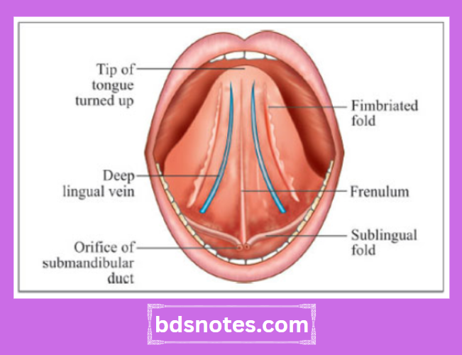
- An inferior surface
- It shows median frenulum linguae
- On either side, there is presence of deep lingual veins
- Oral/anterior 2/3rd
- A curved upper surface/dorsum It is divided into
- It has
- Root:
Posteriormost part of tongue:
- Posteriormost part of tongue is connected to the epiglottis by
- Median glossoepiglotic fold
- Right & left lateral glossoepiglotic fold
- Posteriormost part of tongue separates vallecula which is a depression, from the piriform fossa
Papillae of the tongue:
1. Vallate or circumvallate papillae
- Situated immediately in front of the sulcus terminalis
2. Fungiform papillae
- Present near the tip & margins of the tongue & scattered over the dorsum
3. Filiform papillae
- Covers the presulcal area
4. Foliate papillae
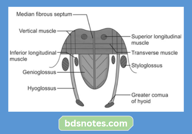
Muscles of tongue:
1. Intrinsic Muscles:
- Superior longitudinal
- Inferior longitudinal
- Transverse
- Vertical
2. Extrinsic muscles:
- Genioglossus
- Hyoglossus
- Styloglossus
- Palatoglossus
Muscles of tongue Blood Supply:
- Arterial Supply:
- Lingual arterybranch of external carotid
- Tonsillarbranch of facial artery
- Ascending pharyngealbranch of external carotid
- Venous Drainage:
- Two venae comitantes
- Accompany the lingual artery
- One vena comitantes
- Accompany hypoglossal nerve
- Deep lingual veinprincipal vein
- These veins unite at the posterior border of the hyoglossus
- Deep lingual veinprincipal vein forms lingual vein
- Deep lingual veinprincipal vein ends in internal jugular vein
- Two venae comitantes
Leave a Reply