• Skip to main content
  • Skip to secondary menu
  • Skip to primary sidebar
  • About Us
  • Terms of Use
  • Privacy Policy
  • Disclaimer
  • Contact Us
  • Sitemap

BDS Notes

BDS notes, Question and Answers

  • Public Health Dentistry
  • Periodontics
  • Pharmacology
  • Pathology
  • Orthodontics
    • Anchorage In Orthodontics
    • Mandibular Growth, Functional Matrix
    • Retention and Relapse
  • General Surgery
    • Cysts: Types, Causes, Symptoms
    • Maxillofacial Fractures, Disorders, and Treatments
    • Lymphatic Disorders
    • Neurological and Facial Disorders
  • Temporal And Infratemporal Regions
    • Spinal and Neuroanatomy
  • Dental Materials
    • Dental Amalgam
Home » Tympanic Membrane: Structure, Blood Supply, And Nerve Supply

Tympanic Membrane: Structure, Blood Supply, And Nerve Supply

February 6, 2026 by Kristensmith Taylor Leave a Comment

Tympanic Membrane: Structure, Blood Supply, And Nerve Supply

Question 1. Tympanic membrane
Answer:

  • It is thin, translucent partition between the external acoustic meatus and middle ear
  • It is placed obliquely at an angle of 55 degrées with the floor of meatus
  • It is composed of
    1. Outer cuticular layer of skin
    2. Middle fibrous layer
    3. Inner mucous layer

Tympanic membrane Blood Supply:

  • Outer surface deep auricular branch of maxillary artery
  • Inner surface anterior tympanic branch of maxillary artery

Tympanic membrane Nerve Supply:

  1. Outer surface
    • Anteroinferior part auriculotemporal nerve
    • Posterosuperior part Auricular branch of vagus nerve
  2. Inner surface
    • Tympanic branch of glossopharyngeal nerve

Question 2. Development of tympanic membrane
Answer:

  • Tubo-tympanic recess develops from the dorsal part of the first pharyngeal pouch
  • Tympanic membrane is formed by apposition of this recess and the first ectodermal cleft
  • These two forms the inner and outer epithelial linings
  • The intervening mesoderm forms the connective tissue basis.

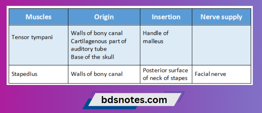
Question 3. Name the muscles of tympanic cavity and their nerve supply.
Answer:

Muscles of tympanic cavity

Muscles Of Tympanic Cavity

Question 4. Internal auditory meatus
Answer:

  • The internal auditory meatus is a canal within the petrous part of the temporal bone of the skull between the posterior cranial fossa and the inner ear.
  • It opens above the anterior part of the jugular foramen
  • It is about 1cm long and runs transversely in a lateral direction
    It is closed Laterally by a perforated plate of bone known as lamina cribosa which separates it from the internal ear.
  • It transmits the seventh and eighth cranial nerves and the labyrinthine vessels.

Filed Under: Anatomy

Reader Interactions

Leave a Reply Cancel reply

Your email address will not be published. Required fields are marked *

Primary Sidebar

Recent Posts

  • Branchial Cleft Cyst: Background, Pathophysiology, Etiology
  • Maxillary Nerve: Origin, Course, And Branches
  • The Father Of Anatomy And A Great Anatomist Herophilus
  • Bone Structure – Anatomy
  • The External Carotid Artery: Anatomy, Branches, And Functions
  • Occipitofrontalis Muscle
  • Superficial Temporal Artery
  • Platysma Muscle
  • Cartilage
  • Cauda Equina And Conus Medullaris Syndromes
  • Subcutaneous Injections And Device Management
  • Types Of Circulation: Pulmonary, Systemic, And Portal
  • Structure Of Skeletal Muscle
  • Elastic Cartilage
  • Cellular Organelles And Structure
  • The Golgi Apparatus – The Cell
  • The Cytoplasmic Inclusions Of Certain Plant Cells
  • Dental Abscess
  • Laser Surgery
  • Our Facial Muscles And Their Functions

Copyright © 2026 · Magazine Pro on Genesis Framework · WordPress · Log in