• Skip to main content
  • Skip to secondary menu
  • Skip to primary sidebar
  • About Us
  • Terms of Use
  • Privacy Policy
  • Disclaimer
  • Contact Us
  • Sitemap

BDS Notes

BDS notes, Question and Answers

  • Public Health Dentistry
  • Periodontics
  • Pharmacology
  • Pathology
  • Orthodontics
    • Anchorage In Orthodontics
    • Mandibular Growth, Functional Matrix
    • Retention and Relapse
  • General Surgery
    • Cysts: Types, Causes, Symptoms
    • Maxillofacial Fractures, Disorders, and Treatments
    • Lymphatic Disorders
    • Neurological and Facial Disorders
  • Temporal And Infratemporal Regions
    • Spinal and Neuroanatomy
  • Dental Materials
    • Dental Amalgam
Home » Middle Ear Anatomy: Medial Wall, Muscles, And Auricle Innervation

Middle Ear Anatomy: Medial Wall, Muscles, And Auricle Innervation

February 6, 2026 by Kristensmith Taylor Leave a Comment

Middle Ear Anatomy: Medial Wall, Muscles, And Auricle Innervation

Question 1. Briefly describe the features of the medial wall of middle ear.
Answer:

Medial wall of middle ear has following features:

1. Promontory

  • It is rounded bulging produced by first turn of the cochlea
  • Grooved by tympanic plexus

2. Fenestra vestibule

  • It is oval opening posteriosuperior to the promontory
  • It leads into the vestibule of the internal ear

3. Prominence of the facial canal

  • It reaches the lower margin of aditus
  • Ends at stylomastoid foramen

4. Fenestra cochlea

  • It is round opening posteroinferior to the promontory
  • It opens into the Scala tympani of the cochlea

5. Sinus tympani

  • It is a depression behind the promontory

6. Processus cochleariformis

  • It is curved lamina between the canals for the tensor tympani and for the auditory tube.

7. Prominence of lateral semicircular canal

  • Present above the facial canal

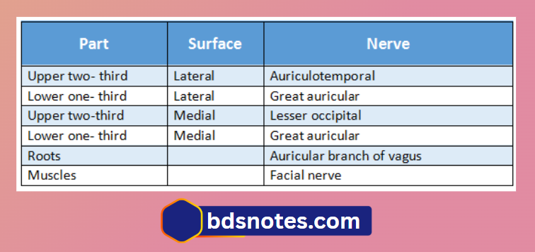
Question 2. Nerve supply to the auricle (pinna)
Answer:

Tensor Tympani And Stapedius

Filed Under: Anatomy

Reader Interactions

Leave a Reply Cancel reply

Your email address will not be published. Required fields are marked *

Primary Sidebar

Recent Posts

  • Branchial Cleft Cyst: Background, Pathophysiology, Etiology
  • Maxillary Nerve: Origin, Course, And Branches
  • The Father Of Anatomy And A Great Anatomist Herophilus
  • Bone Structure – Anatomy
  • The External Carotid Artery: Anatomy, Branches, And Functions
  • Occipitofrontalis Muscle
  • Superficial Temporal Artery
  • Platysma Muscle
  • Cartilage
  • Cauda Equina And Conus Medullaris Syndromes
  • Subcutaneous Injections And Device Management
  • Types Of Circulation: Pulmonary, Systemic, And Portal
  • Hierarchical Organization Of Skeletal Muscle Tissue
  • Elastic Cartilage Histology Short Note For Medical Exams
  • Cellular Organelles And Structure
  • The Golgi Apparatus – The Cell
  • The Cytoplasmic Inclusions Of Certain Plant Cells
  • Dental Abscess
  • Laser Surgery
  • Our Facial Muscles And Their Functions

Copyright © 2026 · Magazine Pro on Genesis Framework · WordPress · Log in