Tonsils: Anatomy, Definition & Function
Question 1. Microscopic anatomy and development of Tonsil
Answer:
- It consists of diffuse lymphoid tissue containing lymphatic nodules
- It is covered by stratified Squamous epithelium
- This epithelium extends into tonsil forming several tonsillar crypts
Microscopic anatomy and development of tonsil Crypts:
- It contains numerous mucous glands
- It contains cells like
- Lymphocytes
- Desquamated epithelium cells
- It also contains bacteria
“Understanding tonsils through FAQs: Anatomy, definition, and functions explained”
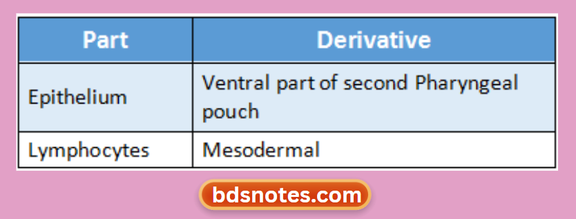
Microscopic anatomy and development of tonsil Development:
“Importance of studying tonsils for medical students: Questions explained”
Question 2. Microscopic structure of Trachea
Answer:
- It is made up of 16-20 tracheal cartilages
- The intervals between the cartilages are filled by fibrous tissue
- Gaps between cartilage end are filled in by smooth muscle & fibrous tissue
- Connective tissue in the wall contains many elastic fibres
“Common challenges in mastering tonsil anatomy notes effectively: FAQs provided”
Microscopic structure of Trachea Lumen:
- Lined by mucous membrane
- It consists of
- Epithelium
- It is pseudostratified ciliated columnar
- Microscopic structure of trachea It contains:
- Goblet cells
- Basal cells
- Lymphocytes
- Connective tissue contains
- Elastic fibres
- Serous & mucous glands
- Lymphoid tissue
- Eosinophil, leucocytes
- Epithelium
Leave a Reply