Understanding Lymph Nodes: Histology, Zones, And Immune Functions
Histology of Lymph Node
Answer:
- They are small beanshaped structures scattered along the course of lymphatic vessels
- Its concavity constitute a hilum for entry & exit of blood vessels
- Histology of medium sized artery It consists of:
- Connective tissue framework
- Interstitial spaces of it is filled with lymphocytes & other cells
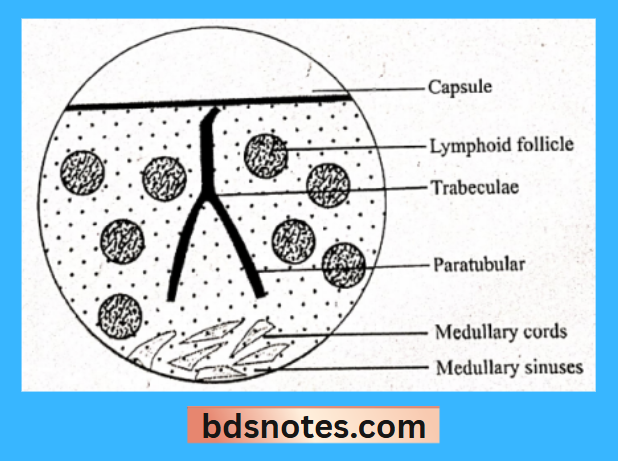
- Lymph node is surrounded by a capsule
- It consists of mainly collagen fibres & some elastic fibres & smooth muscle
- Remaining part of lymph node contains reticular fibres
- Each lymph node is divided into lobules by septa or trabeculae that extendsfrom capsule into the node
- Cells
- Lymphocytes
- Lymphocytes present in blood are of two types
- BLymphocytes
- It divides rapidly
- Matures into plasma cells
- Functions:
- Remain in lymph node as memory cells & produce plasma cells
- Return to blood stream via lymph
- Concern with humoral immunity
- T-Lymphocytes
- Present in between the lymphatic nodules
- Concern with cellmediated immunity
- BLymphocytes
- Lymphocytes present in blood are of two types
- Fibroblasts
- Spindle shaped cell
- Present along reticular fibres
- Macrophages
- Present in lymph sinuses & around germinal center
- More in medulla than in cortex
- Help in phagocytosis
- Endothelial cells
- Present lining the blood vessels of lymph node
- Pericytes & smooth muscle cells
- Present around the blood vessels
- Lymphocytes
- Connective tissue framework
Histology of medium sized artery Zones:
- Cortex
- Outermost zone
- Contains densely packed lymphocytes
- It is darkly stained
- Several rounded areas called lymphatic follicles or nodules are present
- Each nodule contains a germinal center surrounded by densely packed lymphocytes
- Medulla
- It is lighter zone surrounding cortex
- Contains fewer lymphocytes
- These lymphocytes are arranged in the form of branching
2. Histology of medium sized artery Lymphocytes
- Present in the cortex of each lobules as densely packed
- Less densely packed in medulla of lobule
3. Histology of medium sized artery Macrophages
- Placed subjacent to the capsule at corticomedullary junction & in the medulla
Functions: - Phagocytosis
- Deeper cells are dendritic
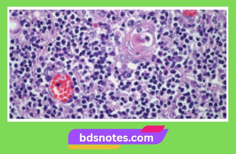
4. Histology of medium sized artery Corpuscles of Hassall
- They are small rounded cells
- Present in medulla
Leave a Reply