Hypocalcification Of Tooth Enamel
Question 1. Hypocalcified structures of enamel.
Answer:
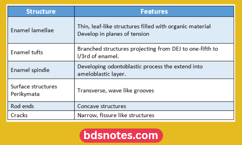
“Hypocalcification Structure Of Enamel And It’s Features”
“Hypocalcification Causes”
Question 2. Enamel knot, cord, and niche
Answer:
Enamel Knot:
- They are clusters of non-dividing epithelial cells present in molar cap stage tooth germ
- It consists of cells that do not divide but promote the division of adjacent epithelial cells
Dental Hypocalcification
- Each tooth germ has a single primary enamel knot at the cap stage which disappears and a secondary knot appears It represents a signaling center for tooth morphogenesis Enamel Cord:
- Temporal nested patterns in the enamel knot that extend between the inner and outer dental epithelium are called enamel cords.
“Hypocalcification And Amelogenesis Imperfecta”
Enamel Niche:
- The Enamel organ appears to have a double attachment to the dental lamina through strands.
- These strands enclose a funnel-shaped depression containing connective tissue. This is known as the enamel niche

“Hypocalcification Symptoms”
Question 3. Enamel cuticle
Answer:
- It is a delicate membrane covering the newly erupted teeth
Enamel Hypocalcification
Enamel cuticle Formation:
- Secreted by ameloblasts when the Enamel organ is retracted from the cervical region Enamel formation is complete
“Hypocalcification Treatment”
Enamel cuticle Functions:
- Protects the surface of enamel from the resorptive activity of adjacent tissue prior to the eruption
Hypocalcification Teeth
Enamel cuticle Fate:
- Removed by mastication
- Replaced by pellicle
Leave a Reply