Turner Syndrome And Developmental Anomalies
Question 1. Turner’s syndrome.
Answer:
Turner’s syndrome. Causes: Monosomy of sex chromosome.
Turner’s syndrome. Effects:
- Agenesis of ovaries
- Mental retardation
- Skeletal abnormalities
- Webbed neck.
- Short stature
- Lymphedema of the extremities
- Broad chest with widely space.
Question 2. Development of pituitary gland/Hypophysis cerebri.
Answer:
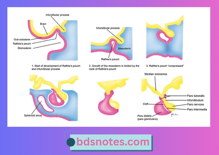
- Pituitary gland develops from the two separate sources.
- The anterior and intermediate parts develop from an ectodermal diverticulum (rathke’s pouch) that grows upwards from the roof of the stomadeum.
- The anterior wall of Rathke’s pouch proliferates to form the pars anterior of the hypophysis.
- The posterior wall remains thin and forms the pars intermedia.
- The pars nervosa and stalk of the hypophysis cereberi develop from a down growth from the floor of the third ventricle in the region of the infundibulum.
- A small extension of this lobe, the pars tuberalis, grows along the stalk of the infundibulum and eventually surrounds it.
- The posterior lobe of the hypophysis is composed from the hypothalamic area.
- The anterior and intermediate parts develop from an ectodermal diverticulum (rathke’s pouch) that grows upwards from the roof of the stomadeum.
Question 3. Development of parathyroid gland.
Answer:
- In the fifth week embryo parathyroid gland develops.
- The inferior parathyroid gland [Parathyroid III] develops from endoderm of the third pharyngeal pouch.
- The superior parathyroid gland [Parathyroid IV] develops from endoderm of the fourth pharyngeal pouch.
- When the thymus descend toward the thorax, parathyroid III is carried along with it.
- But parathyroid IV is prevented from descending as it is closely related to thyroid gland.
- Thus parathyroid III becomes caudal to parathyroid IV.
Leave a Reply