• Skip to main content
  • Skip to secondary menu
  • Skip to primary sidebar
  • About Us
  • Terms of Use
  • Privacy Policy
  • Disclaimer
  • Contact Us
  • Sitemap

BDS Notes

BDS notes, Question and Answers

  • Public Health Dentistry
  • Periodontics
  • Pharmacology
  • Pathology
  • Orthodontics
    • Anchorage In Orthodontics
    • Mandibular Growth, Functional Matrix
    • Retention and Relapse
  • General Surgery
    • Cysts: Types, Causes, Symptoms
    • Maxillofacial Fractures, Disorders, and Treatments
    • Lymphatic Disorders
    • Neurological and Facial Disorders
  • Temporal And Infratemporal Regions
    • Spinal and Neuroanatomy
  • Dental Materials
    • Dental Amalgam
Home » Intraembryonic Mesoderm And Fertilization

Intraembryonic Mesoderm And Fertilization

February 9, 2026 by Kristensmith Taylor Leave a Comment

Intraembryonic Mesoderm And Fertilization

Question 1. Intraembryonic mesoderm.

Answer:

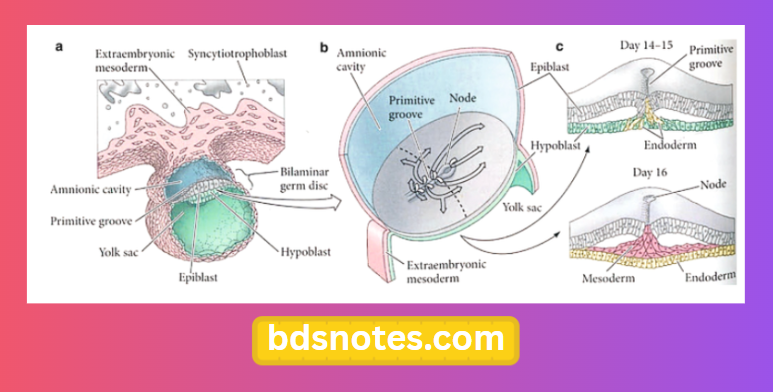
  • The embryonic disc is made up of three germ layers – ectoderm, mesoderm and endoderm.
  • The mesoderm layer present within the embryonic disc is known as intra – embryonic mesoderm.
  • It spreads throughout the disc except in the region of the prochordal plate,
  • It shows three subdivision.
    1. Paraxial mesoderm.
      • It is the mesoderm next to the middle line.
      • It undergoes segmentation to form somites.
    2. Later plate mesoderm.
      • Lateral plate mesoderm.
      • It contains intra-embryonic coelem.
    3. Intermediate mesoderm.
      • It is a strip of mesoderm present between the lateral plate mesoderm and the paraxial mesoderm.

Gastrulation and Germ Layer Development

  • Time of formation: 19 days of IU life.

Question 2. Fertilization
Answer:

  • It is the process by which male and female gametes fuse
  • It occurs in the ampullary region of the uterine tube

Filed Under: Anatomy

Reader Interactions

Leave a Reply Cancel reply

Your email address will not be published. Required fields are marked *

Primary Sidebar

Recent Posts

  • Branchial Cleft Cyst: Background, Pathophysiology, Etiology
  • Maxillary Nerve: Origin, Course, And Branches
  • The Father Of Anatomy And A Great Anatomist Herophilus
  • Bone Structure – Anatomy
  • The External Carotid Artery: Anatomy, Branches, And Functions
  • Occipitofrontalis Muscle
  • Superficial Temporal Artery
  • Platysma Muscle
  • Cartilage
  • Cauda Equina And Conus Medullaris Syndromes
  • Subcutaneous Injections And Device Management
  • Types Of Circulation: Pulmonary, Systemic, And Portal
  • Structure Of Skeletal Muscle
  • Elastic Cartilage
  • Cellular Organelles And Structure
  • The Golgi Apparatus – The Cell
  • The Cytoplasmic Inclusions Of Certain Plant Cells
  • Dental Abscess
  • Laser Surgery
  • Our Facial Muscles And Their Functions

Copyright © 2026 · Magazine Pro on Genesis Framework · WordPress · Log in