• Skip to main content
  • Skip to secondary menu
  • Skip to primary sidebar
  • About Us
  • Terms of Use
  • Privacy Policy
  • Disclaimer
  • Contact Us
  • Sitemap

BDS Notes

BDS notes, Question and Answers

  • Public Health Dentistry
  • Periodontics
  • Pharmacology
  • Pathology
  • Orthodontics
    • Anchorage In Orthodontics
    • Mandibular Growth, Functional Matrix
    • Retention and Relapse
  • General Surgery
    • Cysts: Types, Causes, Symptoms
    • Maxillofacial Fractures, Disorders, and Treatments
    • Lymphatic Disorders
    • Neurological and Facial Disorders
  • Temporal And Infratemporal Regions
    • Spinal and Neuroanatomy
  • Dental Materials
    • Dental Amalgam
Home » Epithelial-Mesenchymal Interaction

Epithelial-Mesenchymal Interaction

July 26, 2025 by Kristensmith Taylor Leave a Comment

Epithelial-Mesenchymal Interaction

Question 1. Epithelial-mesenchymal interaction
Answer:

  • Epithelial-mesenchymal interaction governs the development of epidermal organs like teeth
  • During the early stages of tooth development, a local ectodermal thickening expressing severe signaling molecules appear
  • These signals to the underlying mesenchyme trigger mesenchymal condensation and tooth development

“Factors influencing success with epithelial-mesenchymal interaction studies: Q&A”

Example:

Gene Expression In Tooth Development

“Understanding epithelial-mesenchymal interaction through FAQs: Mechanisms, functions, and uses explained”

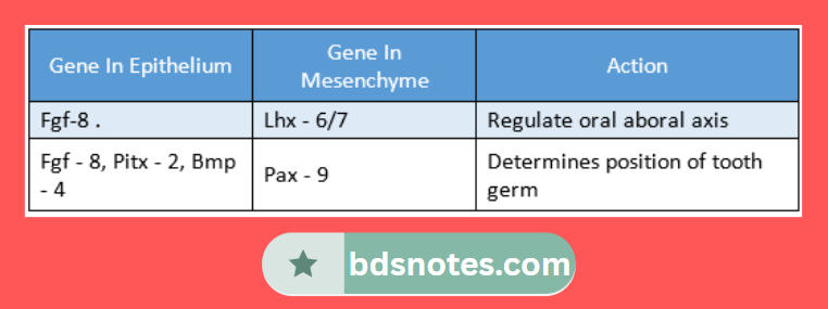
Genes and their action:

  • Pax – 9 – determines morphogenesis
  • LHX – Mesenchymal marker for initiation
  • Fgf – 8 – a marker of the first branchial arch
  • Bmp – 2, Bmp – 4 – patterning of tooth
  • Bmp – 5 – endochondrogenesis
  • Bmp – 7 – dentinogenesis
  • Msx 1, Msx 2 – expresses incisor in mandible
  • Msx 1, Msx 2 &Dlx – 2 – express canine
  • Dlx 2 &Barx 1 – expresses molar

“Importance of studying epithelial-mesenchymal interaction for medical students: Questions explained”

Question 2. Dental lamina.
Answer:

  • It results from the dividing of the primary epithelial band at around the 7th week of intrauterine life.
  • It represents the lingual process of this band.
  • It later breaks to form discrete epithelial islands which are named epithelial pearls.
  • It serves as the primordium for the ectodermal portion of the deciduous teeth,
  • It has a surface of squamous cells and a basal layer of columnar cells.

“Common challenges in mastering epithelial-mesenchymal interaction notes effectively: FAQs provided”

Filed Under: Anatomy

Reader Interactions

Leave a Reply Cancel reply

Your email address will not be published. Required fields are marked *

Primary Sidebar

Recent Posts

  • Branchial Cleft Cyst: Background, Pathophysiology, Etiology
  • Maxillary Nerve: Origin, Course, And Branches
  • The Father Of Anatomy And A Great Anatomist Herophilus
  • Bone Structure – Anatomy
  • The External Carotid Artery: Anatomy, Branches, And Functions
  • Occipitofrontalis Muscle
  • Superficial Temporal Artery
  • Platysma Muscle
  • Cartilage
  • Cauda Equina And Conus Medullaris Syndromes
  • Subcutaneous Injections And Device Management
  • Types Of Circulation: Pulmonary, Systemic, And Portal
  • Structure Of Skeletal Muscle
  • Elastic Cartilage
  • Cellular Organelles And Structure
  • The Golgi Apparatus – The Cell
  • The Cytoplasmic Inclusions Of Certain Plant Cells
  • Dental Abscess
  • Laser Surgery
  • Our Facial Muscles And Their Functions

Copyright © 2026 · Magazine Pro on Genesis Framework · WordPress · Log in