“What are the key differences between cardiac and skeletal muscle? A detailed question and answers guide”
Cardiac Muscle Vs Skeletal Muscle Differences
When we think about muscles, we often picture the biceps or the legs of an athlete. But there are different types of muscle in our bodies, and each has a unique role. Cardiac muscle and skeletal muscle are two key types, and they serve very different purposes. Understanding the difference between cardiac muscle and skeletal muscle can help us appreciate how our bodies function and maintain health.
Cardiac Muscle vs Skeletal Muscle
“Understanding cardiac vs skeletal muscle through FAQs: Anatomy, function, and physiology explained”
- Cardiac muscle is found only in the heart and operates involuntarily, while skeletal muscle is attached to bones and is under voluntary control.
- Both muscle types are striated, meaning they have a striped appearance, but they differ in structure and function.
- Cardiac muscle contracts at a steady pace to pump blood, whereas skeletal muscle can contract quickly for short bursts of energy.
- Skeletal muscles can be fast-twitch or slow-twitch, affecting how they respond to exercise and fatigue.
- Issues with cardiac muscle can lead to heart diseases, while problems with skeletal muscle can result in various muscle disorders.
“Importance of studying cardiac vs skeletal muscle for healthcare professionals: Questions explained”
Skeletal Muscle Action Potential
Key Characteristics Of Cardiac Muscle
Location and Structure
Cardiac muscle, as the name suggests, is exclusively found in the heart. It forms the myocardium, the thick middle layer of the heart wall responsible for the heart’s pumping action. Unlike skeletal muscle, cardiac muscle cells (cardiomyocytes) are shorter and branched. These branches interconnect with each other through specialized junctions called intercalated discs, forming a syncytium, which allows for rapid and coordinated contraction of the heart.
Involuntary Control
“Common challenges in differentiating cardiac vs skeletal muscle effectively: FAQs provided”
One of the defining features of cardiac muscle is its involuntary nature. You don’t consciously control your heartbeat; it’s regulated by the autonomic nervous system and intrinsic factors within the heart itself. This is in contrast to skeletal muscle, which is under voluntary control. The heart’s ability to beat rhythmically without conscious input is due to specialized cells called pacemaker cells, which generate electrical impulses that spread throughout the heart muscle, triggering contraction. This intrinsic rhythmicity ensures continuous and reliable heart function.
Cardiac Muscle Contraction Mechanisms
Striated Appearance
Like skeletal muscle, cardiac muscle exhibits a striated appearance under a microscope. This striation is due to the arrangement of actin and myosin filaments into repeating units called sarcomeres. These sarcomeres are responsible for muscle contraction. The organized arrangement of these filaments gives cardiac muscle its characteristic striped pattern. This structural similarity reflects the shared mechanism of contraction between cardiac and skeletal muscle, both relying on the sliding filament theory. The heart’s pumping action is essential for life.
Cardiac muscle’s unique structure and involuntary control are crucial for its function. The interconnected cells and intrinsic rhythmicity ensure coordinated and continuous heartbeats, essential for circulating blood throughout the body. This specialized tissue is designed for endurance and reliability, reflecting the heart’s vital role in maintaining life.
“Factors influencing success with cardiac vs skeletal muscle knowledge: Q&A”
Key Characteristics Of Skeletal Muscle
Location and Structure
Skeletal muscles are the ones we usually think about when we talk about muscles. They’re attached to our bones by tendons, and they’re what allow us to move. Think about lifting a weight, walking, or even just smiling – that’s all skeletal muscles at work. These muscles are made up of long, cylindrical fibers that are bundled together. They’re pretty big compared to other types of muscle cells.
Heart Muscle vs Skeletal Muscle
Voluntary Control
Unlike cardiac or smooth muscle, you have conscious control over your skeletal muscles. This means you decide when and how to move them. Want to pick up a cup of coffee? Your brain sends a signal to the muscles in your arm, telling them to contract. It’s a pretty amazing system when you think about it.
Skeletal Muscle Vs Cardiac Muscle Structure
Fast-Twitch vs Slow-Twitch
Skeletal muscles aren’t all the same. They have different types of fibers that affect how they function. There are two main types:
- Fast-twitch fibers: These fibers contract quickly and powerfully, but they also fatigue faster. They’re great for short bursts of activity, like sprinting or lifting heavy objects.
- Slow-twitch fibers: These fibers contract more slowly and with less force, but they can keep going for a long time without getting tired. They’re important for endurance activities, like running a marathon or cycling.
- Fiber Type Distribution: The proportion of fast-twitch and slow-twitch fibers varies from person to person and even within different muscles in the same person. Genetics, training, and activity level all play a role in determining fiber type composition.
Understanding the difference between fast-twitch and slow-twitch fibers can be really helpful for athletes and anyone interested in improving their physical performance. By tailoring your training to target specific fiber types, you can optimize your strength, speed, and endurance.
“Steps to explain cardiac muscle characteristics: Striations vs intercalated discs vs involuntary control: Q&A guide”
Functional Differences Between Cardiac Muscle And Skeletal Muscle
Role in the Body
Cardiac and skeletal muscles have very different jobs. Cardiac muscle’s main role is to pump blood throughout the body, and it does this tirelessly, 24/7. Skeletal muscle, on the other hand, is all about movement. Think walking, lifting, or even just maintaining posture. They’re the muscles you consciously control to interact with the world. The cardiac muscle tissue is only found in the heart.
Energy Requirements
Cardiac muscle is a real workhorse. It needs a constant supply of energy to keep the heart beating. It’s super efficient at using oxygen to generate ATP, its primary fuel. Skeletal muscle is more flexible. It can use both aerobic (with oxygen) and anaerobic (without oxygen) metabolism. This means it can handle both long, steady activities and short bursts of intense activity. However, this flexibility comes at a cost: skeletal muscle can fatigue more quickly than cardiac muscle.
Muscle Tissue Differences
Contraction Speed
Cardiac muscle contracts at a moderate pace. It needs to contract long enough to effectively pump blood, but not so fast that the heart can’t fill properly between beats. Skeletal muscle, however, can contract at a wide range of speeds. Fast-twitch fibers are designed for quick, powerful contractions, while slow-twitch fibers are better for endurance activities. This difference in contraction speed allows for a wide variety of movements and activities.
Cardiac muscle relies almost exclusively on aerobic metabolism, making it highly resistant to fatigue but also very sensitive to oxygen deprivation. Skeletal muscle, while capable of anaerobic metabolism, fatigues more readily due to the buildup of lactic acid and other metabolic byproducts.
“Role of intercalated discs in cardiac muscle function: Questions answered”
Cardiac Muscle Proteome
Similarities Between Cardiac Muscle And Skeletal Muscle
Striated Structure
Both cardiac and skeletal muscle tissues share a striated appearance when viewed under a microscope. This is due to the highly organized arrangement of actin and myosin filaments within the sarcomeres. These bands create the characteristic light and dark patterns that give these muscle types their distinctive look. It’s a visual cue that tells us a lot about how these muscles contract and generate force.
Presence of Myofibrils
Both muscle types are packed with myofibrils, which are the basic rod-like units of a muscle cell. These myofibrils are composed of repeating sections called sarcomeres, the functional units responsible for muscle contraction. Each sarcomere contains the proteins actin and myosin, which interact to cause the muscle to shorten. The density and arrangement of myofibrils directly impact the muscle’s ability to generate force. Think of them as the engines within the muscle cells, driving movement and power.
“Early warning signs of gaps in understanding cardiac muscle properties: Common questions”
Calcium Dependency
Calcium ions play a vital role in the contraction process for both cardiac and skeletal muscles. Here’s how it works:
- An action potential triggers the release of calcium ions.
- Calcium binds to troponin, causing a shift in tropomyosin.
- This shift exposes myosin-binding sites on actin filaments.
Without calcium, the interaction between actin and myosin cannot occur, and the muscle cannot contract. This dependency highlights a shared fundamental mechanism in how these two muscle types function. It’s a critical step in the muscular system that allows us to move and our hearts to beat.
Physiological Responses Of Cardiac Muscle
Adaptation to Exercise
Cardiac muscle is pretty amazing when it comes to adapting to exercise. Regular physical activity can lead to some beneficial changes in the heart, making it more efficient. For example, endurance training can increase the heart’s stroke volume, which is the amount of blood pumped with each beat. This means the heart doesn’t have to work as hard to deliver the same amount of oxygen to the body. It’s like upgrading to a more powerful engine that uses less fuel. This adaptation is crucial for maintaining cardiovascular health and improving overall fitness. The heart’s ability to adapt ensures it can meet the increased demands of exercise without undue strain. Over time, this can lead to a lower resting heart rate and improved exercise capacity. Think of it as the heart getting smarter and stronger with each workout.
Skeletal Muscle Hypertrophy
Fatigue Resistance
One of the coolest things about cardiac muscle is its incredible resistance to fatigue. Unlike skeletal muscle, which can tire out pretty quickly during intense activity, cardiac muscle is designed to work continuously without giving up. This is super important because, well, you need your heart to keep beating! This resistance comes down to a few key factors. First, cardiac muscle has a ton of mitochondria, the powerhouses of the cell, which generate the energy needed for contraction. Second, it has a really efficient system for using oxygen. And third, it has a special way of handling calcium, which is essential for muscle contraction. All these things work together to make cardiac muscle incredibly resilient. It’s like having a reliable pump that just keeps going and going, no matter what.
Heart Rate Regulation
Heart rate regulation is a complex process that involves both the nervous system and hormones. The heart has its own internal pacemaker, called the sinoatrial (SA) node, which sets the basic rhythm. However, this rhythm can be adjusted by external factors. The autonomic nervous system, which controls involuntary functions, plays a big role. The sympathetic branch increases heart rate, while the parasympathetic branch decreases it. Hormones like adrenaline can also boost heart rate. Factors like body temperature, stress, and physical activity can all influence heart rate. It’s a delicate balance that ensures the heart is pumping blood at the right speed to meet the body’s needs. Here’s a quick look at some factors:
- Autonomic Nervous System
- Hormonal Influence
- Physical Activity
The heart’s ability to regulate its rate is essential for maintaining blood pressure and ensuring adequate oxygen delivery to tissues. This intricate control system allows the heart to respond quickly to changing demands, whether it’s during exercise, stress, or even just changing positions.
“Asymptomatic vs symptomatic effects of ignoring cardiac muscle health: Q&A”
Physiological Responses Of Skeletal Muscle
Adaptation to Training
Skeletal muscle is pretty amazing because it can change a lot depending on how you train it. This ability to adapt is called plasticity, and it’s how your muscles get stronger or more efficient. For example, endurance training, like running, leads to increased mitochondrial density in muscle cells, which helps them use oxygen better. Strength training, on the other hand, causes muscle fibers to get bigger, leading to increased strength.
Fatigue and Recovery
Ever wondered why your muscles get tired during exercise? It’s a complex process, but it mainly comes down to energy depletion and the buildup of metabolic byproducts, like lactic acid.
- The type of activity affects fatigue differently.
- High-intensity exercise leads to faster fatigue.
- Recovery involves replenishing energy stores and clearing out those metabolic byproducts.
Proper nutrition and rest are key for muscle recovery. Without enough of either, you’re setting yourself up for decreased performance and increased risk of injury.
Skeletal Muscle Vs Cardiac Muscle Physiology
Muscle Hypertrophy
Muscle hypertrophy is just a fancy way of saying muscle growth. It happens when you consistently challenge your muscles with resistance training. The muscle fibers experience micro-tears, which then get repaired and rebuilt, resulting in bigger, stronger muscles. Several factors influence hypertrophy, including genetics, nutrition, and training intensity. Here’s a quick look at how training volume impacts muscle growth:
Clinical Implications Of Cardiac Muscle And Skeletal Muscle
Heart Diseases
When things go wrong with cardiac muscle, the consequences can be severe. Cardiomyopathy, for example, weakens the heart muscle, making it harder to pump blood. Coronary artery disease, where plaque builds up in the arteries, can lead to heart attacks due to lack of oxygen reaching the heart muscle. Arrhythmias, or irregular heartbeats, can also stem from problems within the cardiac muscle’s electrical system. Early diagnosis and management are key to improving outcomes in these conditions.
“Differential applications of cardiac vs smooth muscle: Questions answered”
Muscle Disorders
Skeletal muscle disorders are a diverse group, ranging from genetic conditions like muscular dystrophy, where muscles progressively weaken and waste away, to acquired conditions such as myositis, where inflammation damages muscle tissue. Injuries, like strains and tears, are also common, especially in athletes. Here’s a quick look at some common issues:
- Muscular Dystrophy: Genetic, progressive muscle weakness.
- Myositis: Muscle inflammation, often autoimmune.
- Rhabdomyolysis: Breakdown of muscle tissue, releasing harmful substances into the bloodstream.
Rehabilitation Approaches
Rehabilitation is crucial for both cardiac and skeletal muscle issues, but the approaches differ significantly. Cardiac rehabilitation focuses on improving cardiovascular health through exercise, education, and counseling, helping patients recover after a heart attack or heart surgery. Skeletal muscle rehabilitation, on the other hand, aims to restore strength, flexibility, and function after injury or surgery, often involving physical therapy and targeted exercises. The goal is to get people back to their normal activities, whether it’s returning to work or participating in sports. For example, after a knee replacement, physical therapy is essential to regain full range of motion.
Skeletal Muscle Vs Cardiac Muscle Adaptations
Cardiac Muscle And Skeletal Muscle Conclusion
In summary, understanding the differences between cardiac and skeletal muscles is pretty important. Cardiac muscle keeps your heart pumping without you even thinking about it, while skeletal muscle is what you consciously control to move around. They each have their own unique roles in the body, and knowing how they work can help you appreciate the complexity of our muscular system. So next time you think about muscles, remember that not all of them are the same, and each type plays a vital part in keeping us alive and active.
Cardiac Muscle And Skeletal Muscle Frequently Asked Questions
Question 1. What Is The Main Difference Between Cardiac And Skeletal Muscle?
Answer: Cardiac muscle is found only in the heart and works automatically, while skeletal muscle is attached to bones and you control its movement.
Question 2. Are Cardiac Muscles Voluntary Or Involuntary?
Answer: Cardiac muscles are involuntary, meaning they work without you having to think about it.
Question 3. How Do Skeletal Muscles Differ In Speed?
Answer: Skeletal muscles can be fast-twitch, which contract quickly for short bursts of energy, or slow-twitch, which are used for longer activities.
Question 4. What Do Both Cardiac And Skeletal Muscles Have In Common?
Answer: Both types of muscles have a striated appearance and rely on calcium to help them contract.
“Steps to master cardiac vs skeletal muscle for exams: Study plans vs mock tests: Q&A guide”
Question 5. Can Cardiac Muscle Get Tired?
Answer: Cardiac muscle is designed to resist fatigue and can keep working continuously without tiring.
Question 6. What Happens To Skeletal Muscles When You Exercise?
Answer: When you exercise, skeletal muscles can grow larger and stronger, a process known as hypertrophy.
Cardiac Muscle Vs Skeletal Muscle Biology
Difference Between Cardiac Muscle And Skeletal Muscle
The skeletal muscles are striated and attached to the bones of the skeleton. They are found mainly in the limbs and trunk of the body.
Muscle Tissue Differences
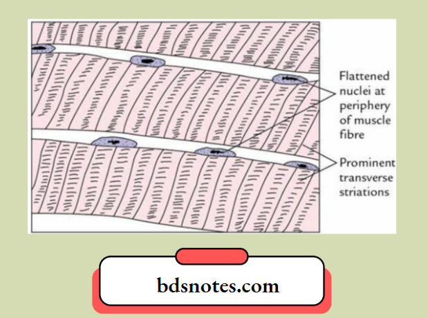
Skeletal Muscles Histological features
- Muscle fibres are elongated, cylindrical and multinucleated.
- Muscle fibres present prominent cross-striations with alternating dark ‘A’ and light ‘I’ bands.
- Nuclei are flat and located at the periphery.
- Muscle fibres do not show branching.
“Role of diagrams in understanding muscle fiber types and structures: Questions answered”
The cardiac muscle is striated, involuntary and located exclusively in the heart.
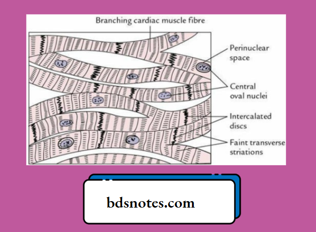
Cardiac Muscle Histological features
- Cardiac muscle fibres are short and thick. They branch and anastomose to form syncytium.
- Cardiac muscle fibres are joined end-to-end at junctional specializations called intercalated discs.
- Each cardiac muscle fibre has a centrally located single oval nucleus.
- Cardiac muscle fibres present faint cross-striations with alternating dark ‘A’ and light ‘I’ bands, i.e. they are not as conspicuous as in skeletal muscle.
“Early warning signs of poor performance in cardiac vs skeletal muscle exams: Common questions”
Leave a Reply