Bitewing Radiography Of Posterior Teeth
Describe The Technique Of Taking Bitewing Radiography Of Posterior Teeth.
Answer. Bitewing radiography is an intraoral radiographic technique.
Bitewing Radiography of Posterior Teeth Principles
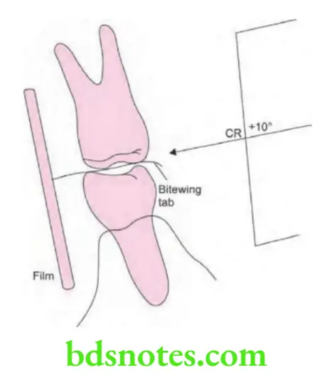
- The film is placed in the mouth parallel to the crown of both the upper and lower teeth.
- The film is stabilized when the patient bites on the bitewing tab or bitewing film holder.
- The central ray of the X-ray beam is directed through the contact area of the teeth, using a + 10° vertical angulation.
“Understanding the role of bitewing radiography in diagnosing dental caries: Q&A explained”
bitewing radiography
“Importance of studying bitewing radiography for better dental diagnostics: Questions explained”
Film holder and Bitewing tab
A film holder is used to stabilize the film. Those used for bite wing radiography are:
- Rinn XCP bite wing instruments: Which have a plastic bite blocks, plastic aiming rings and metal indicator arms. A snap ring collimator may be added to reduce the exposure to the patient. These holders are reusable.
Dental bitewing radiographs
- Bitewing tab: It is an alternative to a film-holding device. A film can be fitted with a bite tab or bite loop. This is a heavy paper board tab or loop with adhesive which may be attached to the white side (tube side) of the periapical film. Readymade bitewing films with attached tabs are also available.
Posterior interproximal radiography
“Common challenges in performing bitewing radiography effectively: FAQs provided”
Position of Patient
- Briefly explain the procedure to patient.
- Ask the patient is seated upright and the chair adjusted to a comfortable working position.
- Adjust the head rest to support and position the patient’s head so that the upper arch is parallel to the floor and the mid sagittal (middle) plane is perpendicular to the floor.
- Secure the lead apron and thyroid collar.
- Remove all foreign objects from the face and mouth.
“Steps to explain different types of bitewing radiographs: Vertical vs horizontal: Q&A guide”
Intraoral bitewing radiography
Five Basic Rules to follow in the Bite Wing technique
- Film placement: The film must be placed to cover the prescribed area.
- Film position: The film must be positioned parallel to the crowns of both the upper and lower teeth and stabilized by biting on the film holder or tab.
- Vertical angulation: The central ray must be directed at +10°.
- Horizontal angulation: The central ray must be directed through the contact areas between the teeth.
- Film exposure: The X-ray beam must be centered on the film to ensure that all the areas of the film are exposed and thus partial image or cone cut is avoided.
Leave a Reply