Periapical Granuloma
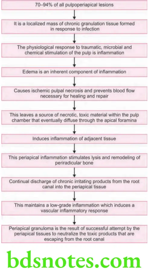
Periapical Granuloma Pathogenesis
Flow chart in next column representing the pathogenesis.
Periapical Granuloma Clinical Features
- Tooth is often asymptomatic.
- Previous history of prolonged sensitivity to heat or cold.
- Deep caries, a deep restoration or history of trauma.
- Tooth is often tender to percussion.
- Fail to react to thermal or electrical stimulation (because the pulp is nonvital).
“Steps to explain clinical features of periapical granuloma: Symptoms vs signs: Q&A guide”
Periapical Granuloma – Clinical Features, Radiographic Signs & Treatment
Periapical Radiolucencies Diagnosis
“Understanding the role of periapical granuloma in endodontics: Q&A explained”
Differential Diagnosis Of Periapical Radiolucencies
Periapical Granuloma Radiographic Features
- Earliest sign: Thickened PDL space at the root apex
- Well-circumscribed radiolucency: Somewhat rounded and surrounding the apex of the tooth
Clinical Features of Periapical Granuloma
- Thin radiopaque hyperostotic border.
Radiographic Features Of Periapical Radiolucencies
Periapical Granuloma Microscopic Features
- Proliferating endothelialcells, capillaries, young firoblasts, collagen, chronic inflammatory cells, lymphocytes, plasma cells, macrophages.
- Nests of odontogenic epithelium, Russell bodies, foam cells, cholesterol cleft.
“Importance of studying periapical granuloma for accurate diagnosis: Questions explained”
Radiographic Appearance of Periapical Granuloma
- More inflammation is seen in center of the lesion.
- At periphery, Fibrosis (healing) is seen.
“Common challenges in diagnosing periapical granuloma: FAQs provided”
Periapical Granuloma Treatment
- Extraction of the involved tooth.
- Root canal therapy (with or without apicoectomy).
- If left untreated, it may undergo transformation to periapical cyst.
Leave a Reply